What is lymph node surgery for melanoma?
There are two types of lymph node surgery for cutaneous melanoma:
- Sentinel lymph node biopsy (SLNB) [1].
- Lymph node dissection or completion lymphadenectomy (CLND) [2].
Sentinel lymph node biopsy
A sentinel lymph node biopsy is used to identify and sample the ‘sentinel’ or first lymph node (or nodes) that potential metastatic melanoma would encounter if present in lymphatic vessels draining the site of the primary melanoma.
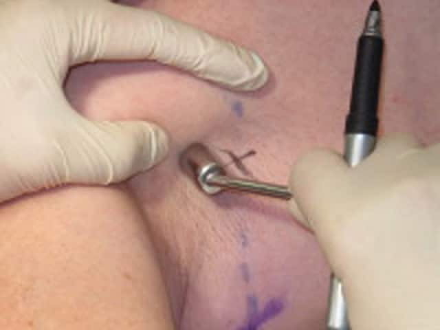
Sentinel lymph node biopsy is performed in people who do not have swollen lymph nodes at the time of re-excision of the biopsy site — known as wide local excision — and is usually under general anaesthetic [3].
A blue dye is injected into the initial biopsy scar prior to the wide local excision. This dye is carried by lymphatic channels to the sentinel lymph node or nodes [3].
- For the upper limb, the sentinel lymph node basin is usually located in the axilla.
- For the lower limb, it is typically found in the inguinal region.
- In-transit sentinel lymph nodes located closer to the melanoma site than the regional nodes have been detected in 10% [4].
- A truncal melanoma can have a sentinel lymph node in the axilla, the inguinal region, or both.
- Lymphatic drainage in the head and neck is unpredictable, with multiple possible locations for sentinel lymph nodes including involvement of multiple basins [5–6].
Lymphoscintigraphy is also used to map the sentinel lymph nodes, using a specialised scanner to detect radiation from a radiotracer combined with the blue dye [7,8].
The sentinel lymph nodes are excised for examination by a pathologist who measures the dimensions of any melanoma found within the lymph nodes and whether it extends beyond the lymph node (which confers poorer prognosis) [9].
Sentinel node biopsy
Lymph node dissection
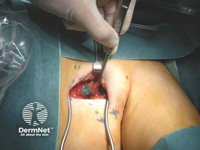
Lymph node dissection or completion lymphadenectomy is the removal of all lymph nodes in the nodal basin (eg, axilla, inguinal region, or head and neck) [2]. This is performed under general anaesthetic. The pathologist examines all the excised lymph nodes for metastatic melanoma [2].
Which patients should be considered for lymph node surgery in melanoma?
Sentinel lymph node biopsy
Whether sentinel lymph node biopsy is performed depends on a melanoma patient’s risk for nodal metastasis. Opinions vary in which patients it should be performed; the key influence was the publication of the ‘MSLT-1’ study [10]. SLNB is currently considered for:
- Cutaneous melanomas that are greater than 0.8 mm in Breslow thickness with an additional adverse prognostic factor (eg, high mitotic rate or ulceration) [10,11].
- Melanomas that are greater than 1 mm in Breslow thickness without other adverse prognostic factors [10,11].
Lymph node dissection
Lymph node dissection is currently considered in two settings:
- If a positive sentinel lymph node involves a large microscopic metastatic melanoma deposit or there are multiple positive lymph nodes
- When there is biopsy-proven lymphadenopathy for metastatic melanoma (ie, the lymph nodes are grossly enlarged) [12].
Melanoma metastases
What are the contraindications with lymph node surgery?
Contraindications mainly relate to sentinel node biopsy.
- Sentinel lymph node biopsy is contraindicated when there has been prior lymph node surgery in the particular basin.
- It should not be performed after wide local excision, flap, or skin graft because its accuracy is reduced [13].
- There is no benefit of performing a sentinel lymph node biopsy if the patient has stage III metastatic disease (palpable involved lymph nodes, satellite/in-transit disease, or distant metastases) [11].
- Older patients or those with significant comorbidities have greater intraoperative risk. A discussion of risk versus benefit is required.
- Patients with pure desmoplastic melanoma have a lower rate of a positive sentinel lymph node biopsy than other subtypes [14].
Tell me more about lymph node surgery
A positive sentinel lymph node biopsy gives prognostic information for risk stratification and staging; it does not have therapeutic benefit. Compared to those who underwent regular observation, those who underwent sentinel lymph node biopsy had no difference in 10-year melanoma-specific survival [10].
Prior to the publication of the results of two studies, ‘DeCOG-SLT’ and ‘MSLT-II’, completion lymph node dissection was recommended for patients with a positive sentinel lymph node biopsy [2,12].
- There was no difference in survival between patients who underwent immediate completion lymph node dissection compared with those had undergone regular clinical and ultrasound surveillance of their lymph node basin with dissection if recurrence was detected [2,12].
- Immediate lymph node dissection offers disease control in the nodal basin, but patients are at risk of surgical complications unlike patients under observation.
- A scoring system to predict non-sentinel lymph node involvement may help select patients who would benefit from immediate completion lymphadenectomy [15].
What are the benefits of lymph node surgery?
Sentinel lymph node biopsy provides prognostic information.
- Negative sentinel lymph node biopsy is reassuring.
- However, a patient with negative sentinel lymph node biopsy may later develop lymph node metastases in that nodal basin [16].
- The results of sentinel lymph node biopsy may enable access to adjuvant immunotherapy, radiotherapy, or clinical trials [17,18].
Completion dissection offers immediate control of nodal metastatic melanoma.
What are the risks/disadvantages of lymph node surgery?
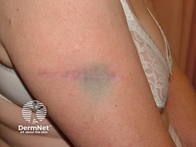
Complication rates for sentinel lymph node biopsy occur in 6–14% of patients [19,20]. The risks of any lymph node surgery include:
- Delayed wound healing from bleeding, infection, or dehiscence
- Poor cosmetic results or discomfort due to tight scarring
- Disruption to the nerve supply in the area
- Seroma due to leaking lymph fluid, with the risk dependent on the number of sentinel lymph nodes removed
- Lymphoedema.
Risks specific to sentinel node biopsy include:
- Anaphylaxis to the blue dye used in the procedure, which occurs in about 1 in 400 patients [19,20].
- Persistent tattoo [20].
- A 5% false-negative risk in the axilla or inguinal lymph node basins [21].
- Up to a 20% false-negative risk in the cervical lymph node basin due to its complex drainage [21].
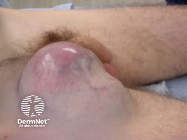
The risks of complications from completion dissection are more common and serious than for sentinel lymph node biopsy, especially lymphoedema, secondary bacterial skin infection, delayed healing, nerve damage, and tight scar formation [22,23].
Lymphoedema is the swelling of the limb distal to the dissected nodal basin due to disruption of lymphatic fluid return. This can be very disabling for patients with only modest improvement achieved from wearing compression garments and massage/exercise [24]. The risk of developing lymphoedema is greater with inguinal completion dissection than with axillary dissection.
Completion dissection may not prevent the development of metastases elsewhere.