

Onychomatricoma — extra information
Introduction Demographics Causes Clinical features Complications Diagnosis Differential diagnoses Treatment Outcome
What is onychomatricoma?
Onychomatricoma is a rare, benign fibroepithelial tumour of the nail matrix [1].
Who gets onychomatricoma?
Onychomatricoma typically affects Caucasian women, with a peak incidence in the fifth decade of life. It is rarely observed in children [2].
What causes onychomatricoma?
The exact pathophysiology of onychomatricoma is unknown, although it could be precipitated by trauma [3,4].
What are the clinical features of onychomatricoma?
Onychomatricoma more commonly affects the fingers than the toes. It could involve either a single digit or multiple digits simultaneously [2,5].
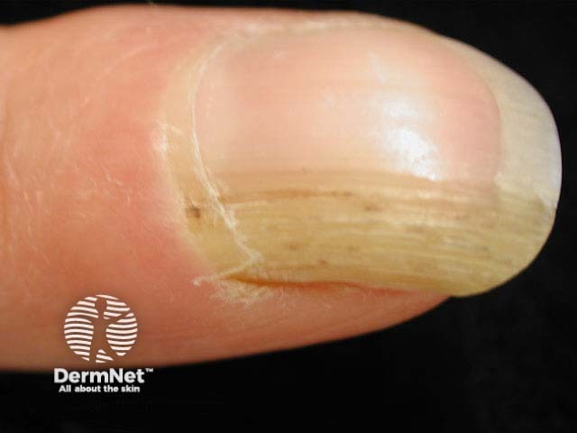
Onychomatricoma is typically slow-growing and painless. Its classic clinical features include [1,4,5]:
- A yellowish and thickened nail plate
- Proximal splinter haemorrhage
- Longitudinal overcurvature of the nail plate
- Finger-like projections penetrating the nail plate, resulting in the characteristic 'woodworm' cavities when viewed from the nail plate’s free margin.
Rarer presentations of onychomatricoma include longitudinal melanonychia, [subungual haematoma]([sitetree_link,id=Loading, please wait...]>), and dorsal pterygium [4,6].
Dermoscopic features of onychomatricoma include perforations in the distal portion of the nail plate, haemorrhagic striae, as well as white longitudinal grooves corresponding to the nail plate channels [5].
What are the complications of onychomatricoma?
Onychomatricoma may be complicated by onychomycosis, so both conditions can coexist [1,5]. Delayed diagnosis and treatment are not uncommon due to unfamiliarity with onychomatricoma [2].
How is onychomatricoma diagnosed?
Onychomatricoma is diagnosed based on its characteristic clinical features supplemented by dermoscopy, imaging, and histopathology [6].
Imaging may be considered to guide clinical decisions before excision if the presentation is non-specific or unclear [4]. Imaging modalities that could help with diagnosing onychomatricoma include [5,7]:
- Ultrasound — this may show hypoechogenic tumour affecting the nail matrix and hyperechogenic area corresponding to the finger-like projections; reduced blood flow could also be observed
- MRI — the affected nail matrix typically shows a low signal uptake, while the finger-like projections have high uptake.
Histopathological evaluation could be performed on either nail clippings or a resected tumour, which is considered the gold standard [1,4–7]:
- Nail clippings — these classically show a thickened nail plate with cavities filled with serous material, lined by a thin layer of epithelium at the periphery
- A resected tumour — distinctive histological features confirm the diagnosis.
- The proximal nail folds — ungual protrusions and deep epithelial invaginations
- The distal zone (corresponding to the lunula) — fibroepithelial projections perforating the nail plate, originating from the matrix.
Immunohistochemistry although helpful, is not routinely required in typical cases [5].
What is the differential diagnosis for onychomatricoma?
Common differential diagnoses of onychomatricoma include:
- Fibrokeratoma (a rare neoplasm)
- Periungual fibroma
- Onychomycosis
- Squamous cell carcinoma
- Intraepidermal squamous cell carcinoma
- Subungual viral wart.
What is the treatment for onychomatricoma?
The definitive treatment for onychomatricoma is complete surgical excision [5].
What is the outcome for onychomatricoma?
Onychomatricoma typically resolves without local recurrence with complete surgical excision [2,4].
References
- Rushing CJ, Ivankiv Roman, Bullock NM, Rogers DE, Spinner SM. Onychomatricoma: a rare and potentially underreported tumour of the nail matrix. Foot Ankle Surg 2017; 56: 1095–98. PubMed
- Chiacchio ND, Tavares GT, Tosti A, et al. Onychomatricoma: epidemiological and clinical findings in a large case series of 30 cases. Br J Dermatol 2015; 173: 1305–07. DOI: 10.1111/bjd.13900. PubMed
- Fierro-Arias L, Corrales-Rosas B, Mercadillo-Pérez P, Medina-Castillo D, Peniche-Castellanos A. Giant onychomatricoma in third toe: exceptional condition with surgical resolution. J Eur Acad Dermatol Venereol 2016; 30: 525–7. DOI: 10.1111/jdv.12923. PubMed
- Cloetingh D, Helm KF, Ioffreda MD, Billingsley E, Rubin AI, Haneke E. JAAD grand rounds quiz. Onychomatricoma. J Am Acad Dermatol 2014; 70: 395–7. DOI: 10.1016/j.jaad.2012.03.029. PubMed
- Joo HJ, Kim MR, Cho BK, Yoo G, Park HJ. Onychomatricoma: a rare tumor of nail matrix. Ann Dermatol 2016; 28: 237–41. DOI: 10.5021/ad.2016.28.2.237. PubMed
- Ocampo-Garza J, Chiacchio NGD, Chiacchio ND. Pigmented onychomatricoma: four cases. Australas J Dermatol 2018; 59: e66–e69. DOI: 10.1111/ajd.12638. Journal
- Tavares GT, Chiacchio NGD, Chiacchio ND, de Souza MV. Onychomatricoma: a tumor unknown to dermatologists. An Bras Dermatol 2015; 90: 265–7. DOI: 10.1590/abd1806-4841.20153650. PubMed
On DermNet