

Atypical solar lentigo — extra information
Introduction Demographics Causes Clinical features Differential diagnoses Complications Diagnosis Treatment Outcome
What is an atypical solar lentigo?
An atypical solar lentigo is a solar lentigo with unusual characteristics. The term may be used when a clinician is unsure of whether a flat brown mark (a lentigo) is a benign solar lentigo or melanoma in situ (an early form of melanoma, a type of skin cancer). The plural of lentigo is lentigines.
Byrom et al gave the name unstable solar lentigo to a specific kind of atypical solar lentigo in sun-damaged skin [1]. Unlike the usual solar lentigo, which is predominantly keratinocytic, unstable lentigo has areas of melanocytic proliferation on histology.
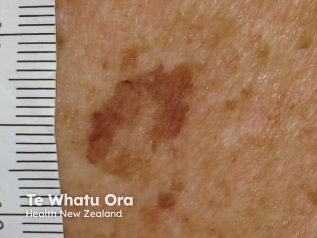
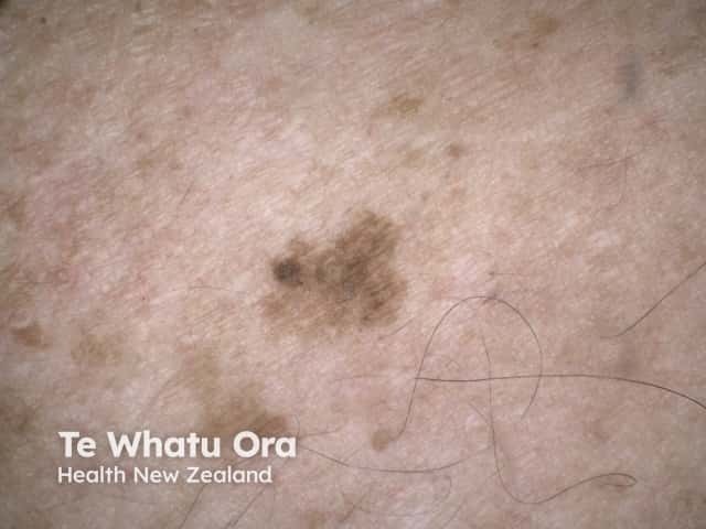
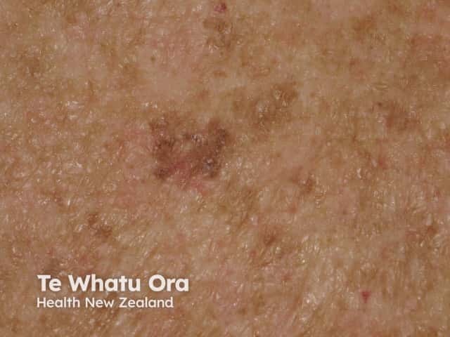
Atypical solar lentigines are large flat brown marks with irregular shape, structure, and colour.
Who gets atypical solar lentigines?
Typical and atypical solar lentigines arise in sun-damaged, ageing skin. Lentigines most commonly affect people over 50 years with fair skin (Fitzpatrick skin type I and II) who spend a lot of time doing outdoor work or outdoor recreation.
Atypical solar lentigines may also arise in people with genetic disorders in which sun damage occurs at an early age (eg, xeroderma pigmentosum), or who are on immunosuppressive medications.
Other features of sun damage may be present in the surrounding, mottled skin [2]. These can include:
- Freckles (ephelides)
- Guttate hypomelanosis
- Solar elastosis
- Actinic keratosis
- Solar lentigo
- Other forms of lentigo
- Seborrhoeic keratosis
- Skin cancer (eg, basal cell carcinoma, squamous cell carcinoma, and melanoma).
What is the cause of atypical solar lentigines?
Atypical solar lentigines are thought to be caused by genetic changes in keratinocytes and in melanocytes due to exposure to ultraviolet radiation. A lichenoid keratosis is due to a local immune reaction.
What are the clinical features of an atypical solar lentigo?
An atypical solar lentigo arises on the face, ears, neck, hands, forearms, or upper back. It is a solitary and distinct macule.
When compared to surrounding solar lentigines, an atypical solar lentigo may:
- Be larger in size and more irregular in shape
- Have a sharper or less distinct margin
- Be darker in colour and have a greater variation in colours
- Be inflamed (red) or there may be areas of hypopigmentation (loss of colour)
- Have greyish or purple discolouration due to an inflammatory reaction.
An atypical solar lentigo may be adjacent to, or form part of, a more typical solar lentigo, a melanoma in situ (whether a lentigo maligna or lentiginous melanoma), or both.
What is the differential diagnosis of an atypical solar lentigo?
The differential diagnoses for an atypical solar lentigo can include:
- Solar lentigo
- Melanoma in situ
- Lichenoid keratosis
- Seborrhoeic keratosis
- Pigmented actinic keratosis
- Other forms of lentigo
- Junctional melanocytic naevus.
What are the complications of an atypical solar lentigo?
The main complication of an atypical solar lentigo is missing a diagnosis of melanoma in situ. Some atypical solar lentigines may also be precursors to melanoma in situ, especially the histological variant, unstable solar lentigo.
How is an atypical solar lentigo diagnosed?
An atypical solar lentigo is evaluated clinically and by dermatoscopy. Pathological examination often leads to a more precise diagnosis, such as solar lentigo, lichenoid keratosis, unstable solar lentigo, atypical lentiginous hyperplasia, atypical junctional melanocytic naevus, or melanoma in situ.
If there is melanocytic proliferation within an atypical solar lentigo, the whole lesion should be excised and multiple layers of the specimens should be examined by histology. Note that a single atypical solar lentigo may show characteristics of solar lentigo, unstable solar lentigo, actinic keratosis, and melanoma in situ (and even invasive melanoma).
What is the treatment for an atypical solar lentigo?
An atypical solar lentigo is often excised.
Lesions that are not excised should undergo a careful follow-up, preferably using serial sequential dermoscopy (mole mapping). Observed changes may include an increase in size, and a change in shape, structure, and colour.
Greater asymmetry in the distribution of structures and colours on dermoscopy should lead to excision of the lesion. Stable lesions can continue to be observed.
What is the outcome for atypical solar lentigo?
The outcome depends on the actual diagnosis.
Solar lentigo and lichenoid keratosis are harmless. Melanoma in situ can progress to invasive melanoma.
References
- Byrom L, Barksdale S, Weedon D, Muir J. Unstable solar lentigo: A defined separate entity. Australas J Dermatol. 2016; 57: 229–34. DOI: 10.1111/ajd.12447. PubMed
- Kossard S. Atypical lentiginous junctional naevi of the elderly and melanoma. Australas J Dermatol 2002; 43: 93–101. PubMed
On DermNet
Other websites
- Lentigo — Medscape