DermNet provides Google Translate, a free machine translation service. Note that this may not provide an exact translation in all languages

Translate

Juvenile dermatomyositis

Author: Dr Hellen Geros, Hospital Medical Officer (PGY2), Western Health, Melbourne, Australia. Copy edited by Gus Mitchell. November 2020.


toc-icon

What is juvenile dermatomyositis?

Juvenile dermatomyositis is the most common form of idiopathic inflammatory myopathy affecting children and adolescents under the age of 18 years.

Who gets juvenile dermatomyositis?

Juvenile dermatomyositis can affect all races and both sexes, although there is a marked female predominance of 5:1. Unlike adult-onset dermatomyositis, there does not appear to be any racial predilection. The peak age of onset is 5–10 years (median 7.4 years). One-quarter of patients present before the age of 4 years. It affects 2–4 children per million per year.

What causes juvenile dermatomyositis?

Juvenile dermatomyositis is a small-vessel autoimmune vasculopathy in which the immune system attacks blood vessels leading to muscle inflammation. The exact pathogenesis remains unclear; however, the following factors may have a role:

  • Genetic susceptibility
  • Environmental factors, such as tobacco smoke and air pollution
  • Infectious triggers, such as viral infections.

There are very few case reports of drug-induced dermatomyositis in children. Malignancy is not associated with juvenile dermatomyositis, unlike adult-onset disease.

What are the clinical features of juvenile dermatomyositis?

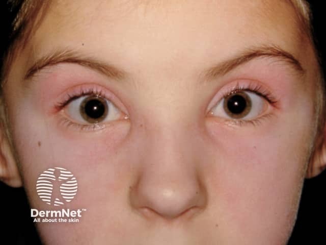
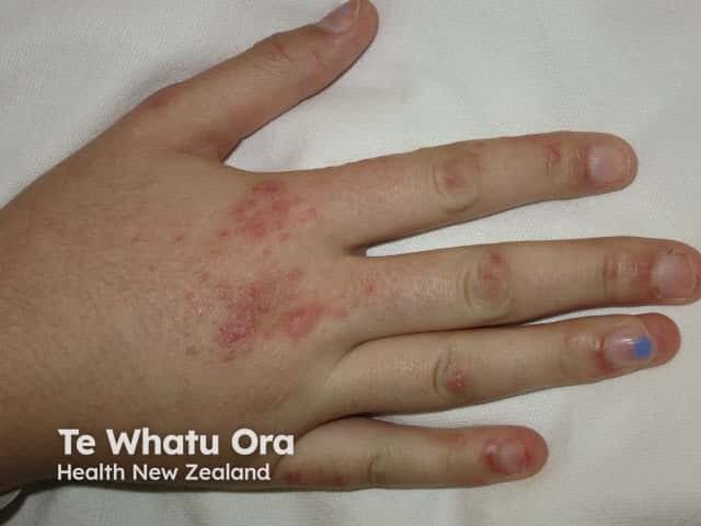
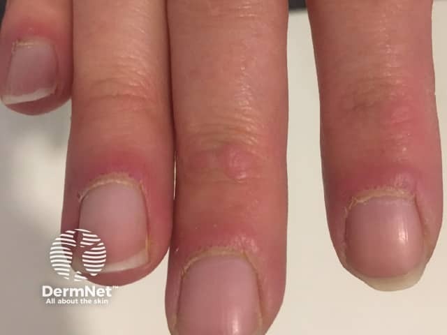
The clinical features of juvenile dermatomyositis are similar to those of adult-onset dermatomyositis, with skin changes often preceding the muscle weakness.

Cutaneous features of juvenile dermatomyositis

  • Heliotrope rash — lilac discolouration around the eyes
  • Gottron papules and/or Gottron sign — purple papules and plaques over the small joints in the hands, elbows, and knees
  • Shawl and/or V sign, and other photosensitive rashes such as malar erythema
  • Dilated or tortuous capillaries in the proximal nailfolds best seen on dermoscopy
  • Skin ulceration affects 20% due to vasculopathy in the skin.

Cutaneous signs of juvenile dermatomyositis

Myositis of juvenile dermatomyositis

  • Symmetrical proximal muscle weakness (thighs, upper arms)
  • Gowers sign describes the use of hands and arms to ‘walk’ up the legs in getting up from a squatting position
  • Dysphagia and voice hoarseness
  • Muscle tenderness, aching, or pain.

Gowers sign
 

What are the complications of juvenile dermatomyositis?

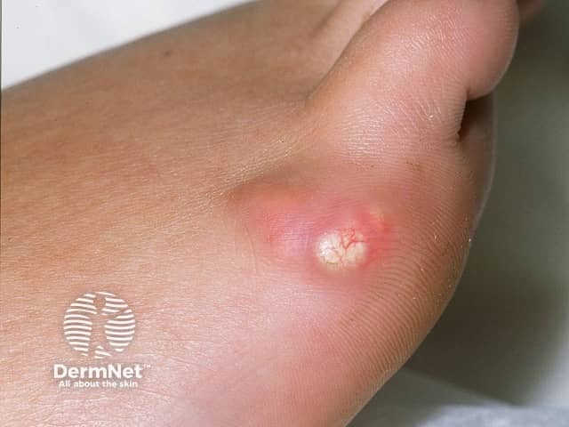
Calcinosis is more common in children with dermatomyositis than in adults, affecting up to 40%. It is a dystrophic calcification in muscle, fascia, subcutaneous tissue, or skin presenting 1–3 years after disease onset. Calcinosis cutis over the elbows, knees, trunk, hands, feet, buttocks, and head presents as hard yellow or white nodules with subsequent ulceration. The development of calcinosis is associated with delayed diagnosis and treatment, and severe or prolonged disease activity.

Lipodystrophy may be localised, partial, or widespread, and affects 10% of juvenile dermatomyositis patients, particularly those with a chronic continuous course; lipodystrophy is rare in adult-onset dermatomyositis. The onset is often delayed; in one large study of 28 patients, lipodystrophy began 4.6 years after diagnosis of juvenile dermatomyositis. Joint contractures and calcinosis cutis were found to be strong predictors for the development of lipodystrophy. As the development of lipodystrophy is associated with diabetes mellitus, insulin resistance, hypertriglyceridaemia, and non-alcoholic steatotic hepatitis, careful monitoring is required in these patients.

Arthritis, either primary joint disease or secondary to the muscle weakness, is common.

Interstitial lung disease, cardiac disease, and liver/bowel involvement are rare but serious complications of juvenile dermatomyositis.

Complications of juvenile dermatomyositis

How is juvenile dermatomyositis diagnosed?

Diagnosis of juvenile dermatomyositis is suspected clinically in a child presenting with the classic skin signs of heliotrope rash, Gottron papules/sign, and myositis (Bohan Peters diagnostic criteria).

Investigations, which may include some of the following, will confirm the diagnosis and assist in predicting prognosis.

  • Autoantibody screen: Anti-p155/140 and anti-MJ autoantibodies are more prevalent in children than adults with dermatomyositis. Anti-tRNA synthetase autoantibodies are uncommon in juvenile dermatomyositis in contrast to adult-onset disease.
    • Anti-p155/140 autoantibodies — photosensitive rashes, skin ulceration and oedema, generalised lipodystrophy, chronic course.
    • Anti-MJ autoantibodies — Caucasian children, young onset of disease, significant muscle effects with impaired physical function, frequent hospitalisation, severe disease less likely to achieve remission.
    • Anti-MDA5 autoantibodies — Asian children, aggressive lung disease, oral and cutaneous ulceration, arthritis, mild muscle disease.
    • Anti-Jo-1 autoantibodies — lung disease, arthritis, fevers, Raynaud phenomenon, mechanic’s hands.
  • Skin biopsy.
  • Muscle investigations — muscle enzymes, muscle biopsy, MRI, electromyography.

What is the differential diagnosis for juvenile dermatomyositis?

Skin signs without myositis:

  • Juvenile-onset amyopathic dermatomyositis; defined as typical skin signs of dermatomyositis without muscle involvement for at least six months
  • Psoriasis.

Muscle weakness and/or myositis without skin changes:

What is the treatment for juvenile dermatomyositis?

The aim of treatment in juvenile dermatomyositis is to control disease activity and induce remission, prevent long term organ damage and deformity, and improve function and quality of life. It generally requires a multidisciplinary team including a general practitioner, physiotherapist, dermatologist, and paediatric rheumatologist.

General measures

Specific measures

Initial treatment requires high dose systemic corticosteroids to rapidly reduce skin and muscle inflammation. The addition of steroid-sparing agents, such as methotrexate or ciclosporin, is more effective than prednisone alone, and allows the corticosteroid dose to be weaned quickly, minimising side effects associated with long term/high dose use. Rituximab has been reported to improve some cutaneous features and myositis in juvenile dermatomyositis; calcinosis, alopecia, poikiloderma, and lipodystrophy showed no major improvements in a large trial of 48 patients. No treatment has been found to consistently improve the calcinosis.

What is the outcome for juvenile dermatomyositis?

The prognosis for juvenile dermatomyositis has markedly improved since the early use of high dose steroids has become the standard of care.

The disease course in one-third is monocyclic (treatment ceased within 2 years with longterm remission), one-quarter is polycyclic (treatment required again after a remission), and the remainder follow a chronic course unable to cease treatment.

With adequate treatment some children lead normal lives with full recovery. However, a significant proportion have a prolonged course with residual muscle weakness and atrophy, joint contractures, and calcinosis. Irreversible muscle damage and calcinosis cutis can significantly impact physical function and quality of life, necessitating assistance with self-care or being unable to walk unassisted.

The mortality rate is 2–3%, with death due to pulmonary, gastrointestinal, cardiac, and multisystem failure.


Bibliography

  • Aggarwal R, Loganathan P, Koontz D, Qi Z, Reed AM, Oddis CV. Cutaneous improvement in refractory adult and juvenile dermatomyositis after treatment with rituximab. Rheumatology (Oxford). 2017;56(2):247–54. doi:10.1093/rheumatology/kew396. PubMed
  • Bingham A, Mamyrova G, Rother KI, et al. Predictors of acquired lipodystrophy in juvenile-onset dermatomyositis and a gradient of severity. Medicine (Baltimore). 2008;87(2):70–86. doi:10.1097/MD.0b013e31816bc604. PubMed Central
  • Hoeltzel MF, Oberle EJ, Robinson AB, Agarwal A, Rider LG. The presentation, assessment, pathogenesis, and treatment of calcinosis in juvenile dermatomyositis. Curr Rheumatol Rep. 2014;16(12):467. doi:10.1007/s11926-014-0467-y. PubMed
  • Huber A, Feldman BM. Long-term outcomes in juvenile dermatomyositis: how did we get here and where are we going? Curr Rheumatol Rep. 2005;7(6):441–6. doi:10.1007/s11926-005-0048-1. PubMed
  • Lundberg IE, Tjärnlund A, Bottai M, et al. 2017 European League Against Rheumatism/American College of Rheumatology classification criteria for adult and juvenile idiopathic inflammatory myopathies and their major subgroups [published correction appears in Ann Rheum Dis. 2018 Sep;77(9):e64]. Ann Rheum Dis. 2017;76(12):1955–64. doi:10.1136/annrheumdis-2017-211468. PubMed
  • Orione MA, Silva CA, Sallum AM, et al. Risk factors for juvenile dermatomyositis: exposure to tobacco and air pollutants during pregnancy. Arthritis Care Res (Hoboken). 2014;66(10):1571–5. doi:10.1002/acr.22358. PubMed
  • Rider LG, Nistala K. The juvenile idiopathic inflammatory myopathies: pathogenesis, clinical and autoantibody phenotypes, and outcomes. J Intern Med. 2016;280(1):24–38. doi:10.1111/joim.12444. PubMed
  • Ruperto N, Pistorio A, Oliveira S, et al. Prednisone versus prednisone plus ciclosporin versus prednisone plus methotrexate in new-onset juvenile dermatomyositis: a randomised trial. Lancet. 2016;387(10019):671–8. doi:10.1016/S0140-6736(15)01021-1. PubMed
  • Wu JQ, Lu MP, Reed AM. Juvenile dermatomyositis: advances in clinical presentation, myositis-specific antibodies and treatment. World J Pediatr. 2020;16(1):31–43. doi:10.1007/s12519-019-00313-8. PubMed

On DermNet

Other websites

Books about skin diseases


Related information

Sign up to the newsletter