Introduction
Demographics
Causes
Clinical features
Variation in skin types
Complications
Diagnosis
Differential diagnoses
Treatment
Prevention
Outcome
Pleomorphic dermal sarcoma (PDS) is a rare dermal-based malignant tumour that typically occurs on sun-damaged skin of the head or neck of elderly patients. Aggressive surgical treatment and close clinical follow-up are required due to high rates of local recurrence and distant metastasis.
PDS shares a clinicopathological overlap with atypical fibroxanthoma (AFX), from which it is separated histologically.

Pleomorphic dermal sarcoma is almost exclusively a tumour that develops in elderly patients with pale, sun-damaged skin and Fitzpatrick skin phototypes 1–3. Patients often have a history of previous skin cancers related to sun damage.
PDS is more common in male and immunosuppressed patients with a median age of 78 years at diagnosis.
PDS has been reported to be caused by ultraviolet (UV) radiation based on its clinical features and genetic studies which have found highly-mutated tumours with recurrent mutations in FAT1, NOTCH1/2, CDKN2A, TP53, and the TERT promoter genes.
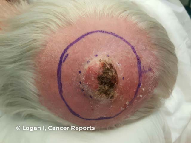
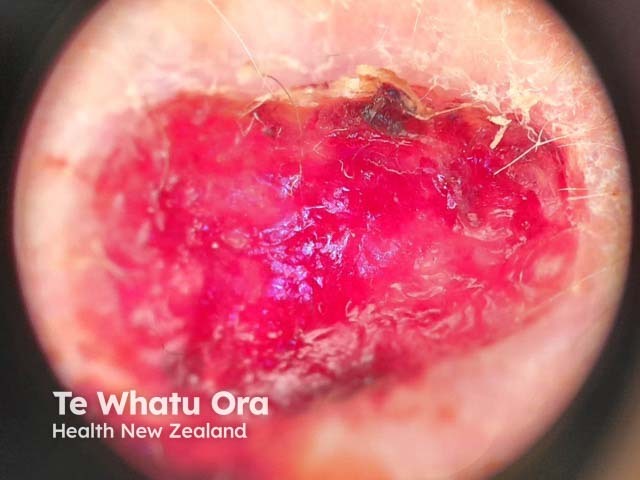
Pleomorphic dermal sarcoma has the following characteristic features:
Sun damage to the adjacent skin is commonly present.
Very rarely, PDS may occur at the site of a previously excised atypical fibroxanthoma as a recurrence, or with distant metastasis at the first clinical presentation.
PDS is a sun damage-related skin malignancy and is not usually found in patients with Fitzpatrick skin types IV–VI. PDS presents in a similar pattern in patients with skin types I–III as the tumour is not pigmented.
Pleomorphic dermal sarcoma is diagnosed only following microscopic assessment. An opinion from a specialist sarcoma histopathologist is recommended for confirmation following excision of the whole tumour.
PDS tumours are composed of atypical spindled and pleomorphic cells originating within the dermis with a sheet-like and fascicular growth pattern with atypical mitotic activity. PDS is excluded from other atypical spindle cell tumours by immunohistochemistry stains.
PDS is morphologically similar to atypical fibroxanthoma from which it is separated by the presence of one or more of the following histologic features:
PDS is not diagnosed clinically. The clinical differential diagnosis is usually:
The histologic differential diagnosis for PDS includes:
The primary treatment for PDS is surgical excision of the tumour, usually with a wide margin of 1–2 cm. Mohs micrographic surgery has also been used and may be considered for sites where tissue conservation is a priority. Large defects may require repair with a skin graft, or dermal matrix substitute.
Due to the risk of local recurrence and metastasis, clinical and radiologic follow-up surveillance has been recommended for a minimum of 4 years following diagnosis.
Local recurrences and metastasis may be managed by:
PDS may be prevented by taking sun protection measures throughout life to minimise photodamage to the skin. This should include covering the head, and in particular the scalp for patients with alopecia, with a wide-brimmed hat or similar sun protective clothing.
The outcome for the majority of patients with PDS is good following complete surgical removal of the tumour.
Between 7–35% of cases have been reported to recur locally. Local recurrence is more common following excisions with narrow or involved pathologic margins, and usually occurs within 1 year of treatment.
Distant metastasis has been reported to occur in 16% of patients in one of the largest cohort studies to date. Distant metastasis is more common in patients who have local recurrence and usually occurs within 3 years of diagnosis.

