DermNet provides Google Translate, a free machine translation service. Note that this may not provide an exact translation in all languages

Translate

Lipodermatosclerosis

Author: Dr Millicent Osti, Resident Medical Officer, The Royal Melbourne Hospital, Melbourne, VIC, Australia; A/Prof Rosemary Nixon, Dermatologist, East Melbourne Dermatology, Melbourne, VIC, Australia. DermNet NZ Editor in Chief: Adjunct A/Prof Amanda Oakley, Dermatologist, Hamilton, New Zealand. Copy edited by Gus Mitchell/Maria McGivern. October 2018.


toc-icon

What is lipodermatosclerosis?

Lipodermatosclerosis is a chronic inflammatory condition characterised by subcutaneous fibrosis and hardening of the skin on the lower legs.

Lipodermatosclerosis is also known as sclerosing panniculitis and hypodermitis sclerodermaformis.

Who gets lipodermatosclerosis?

Lipodermatosclerosis is common, affecting middle-aged or older people. It is more common in women and is associated with immobility and a high body mass index (obesity) [1].

What causes lipodermatosclerosis?

Lipodermatosclerosis is an inflammatory skin condition resulting from underlying venous insufficiency. This is caused by:

  • Incompetent venous valves
  • Venous outflow obstruction
  • Dysfunction of the calf muscle pump [2].

The resulting venous hypertension causes an increase of leukocytes within the veins, which then migrate into surrounding tissue. The leukocytes become activated, attracting and releasing proinflammatory cells and cytokines, inducing a chronic inflammatory state. Increased collagen production leads to the fibrosis of subcutaneous fat [2].

What are the clinical features of lipodermatosclerosis?

Acute phase

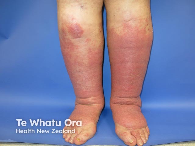
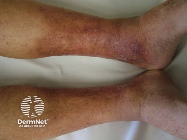
Acute lipodermatosclerosis may mimic cellulitis, with induration, erythema, pain, itch, aching, and a feeling of swelling or heaviness in one or, more often, both lower limbs [2].

In the acute phase, signs may be localised to a single plaque but are usually more widespread. The most commonly affected areas are the pretibial or medial aspect of the leg.

Acute lipodermatosclerosis

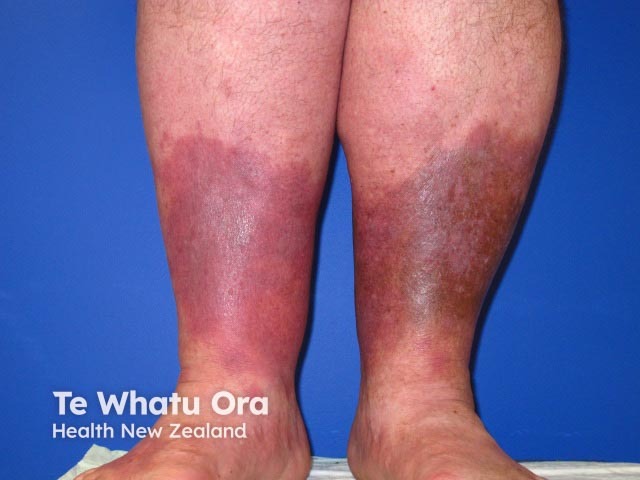
See more images of acute lipodermatosclerosis.

Chronic phase

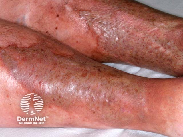
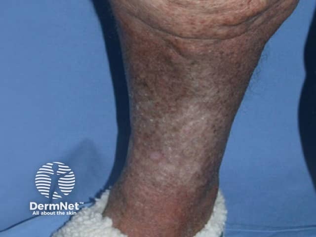
Induration, erythema, and pain continue in the chronic phase of lipodermatosclerosis. Subcutaneous fibrosis may result in significant narrowing of the distal lower limb, causing the leg to have an ‘upside-down champagne bottle’ appearance [1,2].

Other clinical features of chronic venous insufficiency may be present, including hyperpigmentation of the skin from haemosiderin deposition, atrophie blanche, varicose veins, venous eczema, and venous ulcers.

Chronic lipodermatosclerosis

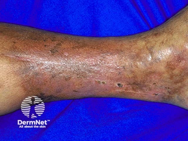
See more images of chronic lipodermatosclerosis.

What are the complications of lipodermatosclerosis?

Lipodermatosclerosis is associated with poor wound healing because of the chronic inflammatory state and fibrosis. Venous ulcers commonly co-exist and may be difficult to treat.

How is lipodermatosclerosis diagnosed?

Lipodermatosclerosis is usually diagnosed clinically. Underlying venous insufficiency may be confirmed using Doppler studies.

biopsy is helpful but should be performed with caution because of the likelihood of poor wound healing [3]. Any biopsy also needs to include the panniculus. The characteristic features seen upon biopsy are dependent on the stage of disease but include subcutaneous lobular and septal changes including:

  • Adipocyte necrosis
  • Pseudocyst formation
  • Lipomembranous (fatty tissue) change
  • Macrophage collections forming lipogranulomas (a nodule of inflamed fat cells around a foreign body)
  • Iron deposition [4,5].

What is the differential diagnosis for lipodermatosclerosis?

Differential diagnoses for lipodermatosclerosis can include:

What is the treatment for lipodermatosclerosis?

General measures

Physical activity (walking) should be encouraged to increase the functionality of the calf muscle pump. Weight reduction is effective if obesity is a factor.

Compression therapy and elevation

Mechanical compression therapy using compression stockings or socks is the mainstay of treatment, encouraging venous return and assisting with symptom control, but may be poorly tolerated in some individuals.

Elevation of the legs can help reduce oedema and pain.

Medical treatment

  • Stanozolol can be effective for pain relief and has also shown to reduce dermal thickness [6].
  • Pentoxifylline has been shown to be useful in venous ulcers in conjunction with compression or in patients who cannot tolerate compression.
  • Combination therapy with hydroxychloroquine may reduce symptoms that are refractory to other treatments [7,8].
  • Intralesional steroid injection of triamcinolone has proven to be effective at alleviating symptoms [9].
  • Tetracyclines, such as doxycycline or minocycline, have anti-inflammatory and anti-angiogenic properties that may provide benefit [10–12].
  • Phlebotonic drugs that modify the tone of the vein wall, including diosmin, hydroxyethylrutoside, or horse chestnut seed extract (escin), may reduce oedema and other symptoms [13].
  • Ultrasound therapy may offer symptomatic benefit, relief of erythema, hardness and pain [14, 15].
  • Ultraviolet radiation (UVA1) has been reported to be effective [16].
  • Emollients and topical steroids are useful in the management of associated venous eczema.

Surgical treatment

Treatment of underlying venous insufficiency can improve symptoms and may reduce the risk of ulcer recurrence. Referral to a vascular surgeon should be considered. Leg vein therapies include:

  • Endovenous ablation by laser, radiofrequency (RF) catheter, steam, cyanoacrylate and mechanical occlusion with chemical assistance (MOCA)
  • Sclerotherapy
  • Vein surgery [17].

What is the outcome for lipodermatosclerosis?

Venous insufficiency is a progressive disease. Although treatment can improve symptoms or slow progression, its associated conditions such as lipodermatosclerosis are usually chronic and recurring.


References

  1. Bruce AJ, Bennett DD, Lohse CM, Rooke TW, Davis MDP. Lipodermatosclerosis: review of cases evaluated at Mayo Clinic. J Am Acad Dermatol 2002; 46: 187–92. PubMed
  2. Bergan JJ, Schmid-Schonbein GW, Smith PD, Nicolaides AN, Boisseau MR, Eklof B. Chronic venous disease. N Engl J Med 2006; 355: 488–98. DOI: 10.1056/NEJMra055289. PubMed
  3. Kirsner RS, Pardes JB, Eaglstein WH, Falanga V. The clinical spectrum of lipodermatosclerosis. J Am Acad Dermatol 1993; 28: 623–7. PubMed
  4. Walsh SN, Santa Cruz DJ. Lipodermatosclerosis: a clinicopathological study of 25 cases. J Am Acad Dermatol 2010; 62: 1005–12. DOI: 10.1016/j.jaad.2009.08.006. PubMed
  5. Choonhakarn C, Chaowattanapanit S, Julanon N. Lipodermatosclerosis: a clinicopathologic correlation. Int J Dermatol 2016; 55: 303–8. PubMed
  6. Dakovic Z, Vesic S, Vukovic J, Pavlovic LJ, Medenica MD. Acute lipodermatosclerosis: an open clinical trial of stanozolol in patients unable to sustain compression therapy. Dermatol Online J 2008; 14(2): 1. PubMed
  7. Jull AB, Arroll B, Parag V, Waters J. Pentoxifylline for treating venous leg ulcers. Cochrane Database Syst Rev 2012; 12: CD001733. DOI: 10.1002/14651858.CD001733.pub3. PubMed
  8. Choonhakarn C, Chaowattanapanit S. Lipodermatosclerosis: improvement noted with hydroxychloroquine and pentoxifylline. J Am Acad Dermatol 2012; 66: 1013–4. DOI: 10.1016/j.jaad.2011.11.942. PubMed
  9. Campbell LB, Miller OF, 3rd. Intralesional triamcinolone in the management of lipodermatosclerosis. J Am Acad Dermatol 2006; 55: 166–8. DOI: 10.1016/j.jaad.2005.09.043. PubMed
  10. Plewig G, Schopf E. Anti-inflammatory effects of antimicrobial agents: an in vivo study. J Invest Dermatol 1975; 65: 532–6. PubMed
  11. Gilbertson-Beadling S, Powers EA, Stamp-Cole M, et al. The tetracycline analogs minocycline and doxycycline inhibit angiogenesis in vitro by a non-metalloproteinase-dependent mechanism. Cancer Chemother Pharmacol 1995; 36: 418–24. DOI: 10.1007/BF00686191. PubMed
  12. Mackelfresh J, Soon S, Arbiser JL. Combination therapy of doxycycline and topical tacrolimus for venous ulcers. Arch Dermatol 2005; 141: 1476–7. DOI: 10.1001/archderm.141.11.1476. PubMed
  13. Martinez-Zapata MJ, Vernooij RW, Uriona Tuma SM, et al. Phlebotonics for venous insufficiency. Cochrane Database Syst Rev 2016; 4: CD003229. DOI: 10.1002/14651858.CD003229.pub3. PubMed
  14. Damian DL, Yiasemides E, Gupta S, Armour K. Ultrasound therapy for lipodermatosclerosis. Arch Dermatol 2009; 145: 330–2. DOI: 10.1001/archdermatol.2009.24. Journal
  15. Hamid SA, Ibrahim ZM,  Mohamady AM. The response of skin hardness and pain sensation to ultrasonic treatment in lipodermatosclerosis patients. Egyptian Journal of Medical Human Genetics, 2014;15: 193-198. Journal.
  16. Patel N, Dahl M. Treatment of lipodermatosclerosis with UVA-1. J Am Acad Derm 2016;74 (5): AB229. Journal.
  17. Gohel MS, Barwell JR, Taylor M, et al. Long term results of compression therapy alone versus compression plus surgery in chronic venous ulceration (ESCHAR): randomised controlled trial. BMJ 2007; 335: 83. DOI: 10.1136/bmj.39216.542442.BE. PubMed

On DermNet

Other websites

Books about skin diseases


Related information

Sign up to the newsletter