Introduction
Demographics
Causes
Demographics at risk
Signs and symptoms
Treatment
Prevention
A leg ulcer is a full thickness skin loss on the leg or foot due to any cause. Leg ulcer occurs in association with a range of disease processes, most commonly with arterial, vascular or neuropathic diseases. A leg ulcer may be acute or chronic.
Chronic leg ulceration affects about 1% of the middle-aged and elderly population. It most commonly occurs after a minor injury in association with:
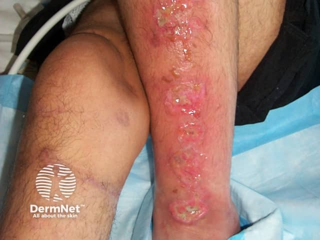
Chronic leg ulcers may also be due to skin cancer, which may be diagnosed by a skin biopsy of the edge of a suspicious lesion. There are also many less common causes of ulcers including systemic diseases such as systemic sclerosis and vasculitis, various skin conditions especially pyoderma gangrenosum, and other causes such as xylazine injection.
An ulcer may be provoked by injury or pressure such as from a plaster cast or ill-fitting ski boot. They may also be caused by bacterial infection, especially impetigo, ecthyma and cellulitis and less often tropical ulcer, tuberculosis or leprosy.
Venous insufficiency refers to improper functioning of the one-way valves in the veins. Veins in the feet and lower legs drain blood uphill to the heart. Two mechanisms assist this uphill flow, the calf muscle pump which pushes blood towards the heart during exercise, and the one-way valves which prevent the flow of blood back downhill. Reflux through the valves, obstruction of the veins and/or impaired calf pumping action results in pooling of blood around the lower part of the leg to just below the ankle. The increased venous pressure causes fibrin deposits around the capillaries, which then act as a barrier to the flow of oxygen and nutrients to muscle and skin tissue. The death of tissue cells leads to ulceration.
Arterial insufficiency refers to poor blood circulation to the lower leg and foot and is most often due to atherosclerosis. In atherosclerosis, the arteries become narrowed from deposits of fatty substances in the arterial vessel walls, often due to high levels of circulating cholesterol and aggravated by smoking and high blood pressure (hypertension). The arteries fail to deliver oxygen and nutrients to the leg and foot resulting in tissue breakdown.
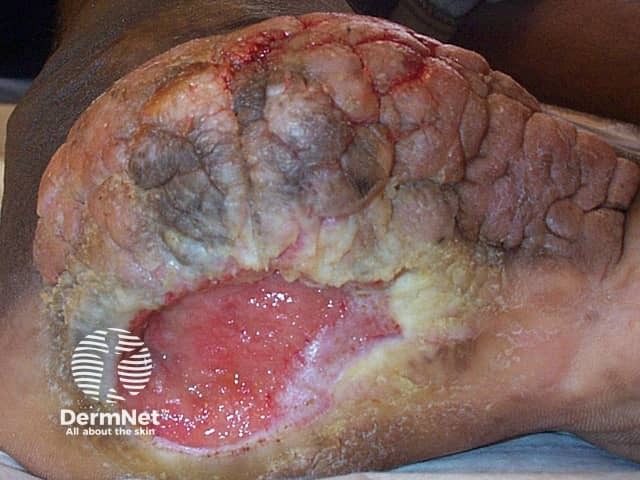
Diabetic ulcers are caused by the combination of arterial occlusion and nerve damage. Although diabetic ulcers may occur on other parts of the body they are more common on the foot. The nerve damage or sensory neuropathy reduces awareness of pressure, heat or injury. Rubbing and pressure on the foot go unnoticed and causes damage to the skin and subsequent ‘neuropathic’ ulceration.
See more about the differential diagnosis of leg ulcers.
Certain conditions have been linked with the development of venous and arterial leg ulcers.
Arterial ulcer
A diabetic ulcer is more likely if diabetes is not well controlled by diet and/or medication. Ulceration is also more likely if there is poor care of the feet, badly fitting shoes, and continued smoking.
The features of venous and arterial ulcers differ somewhat.
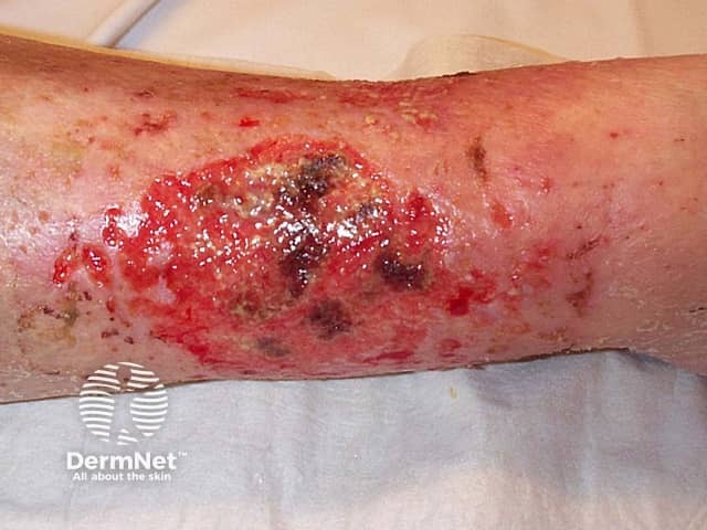
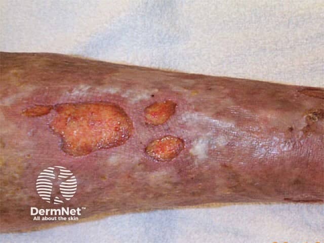
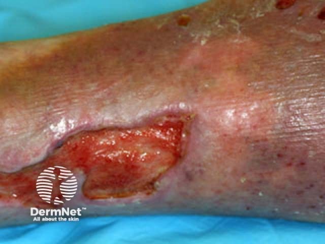
Characteristics of a stasis ulcer include:
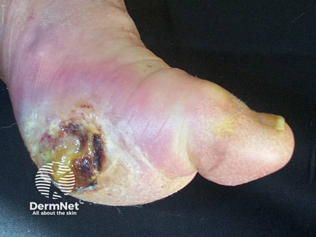
Characteristics of an arterial ulcer include:
A diabetic ulcer has similar characteristics to arterial ulcer but is more notably located over pressure points such as heels, tips of toes, between toes or anywhere the bones may protrude and rub against bed sheets, socks or shoes. In response to pressure, the skin increases in thickness (callus) but with a minor injury, this breaks down and ulcerates.
See more images of leg ulcers.
An infected ulcer characteristically has a yellow surface crust or green/yellow pus and may smell unpleasant. There may be surrounding tender redness, warmth and swelling (cellulitis).
Where possible, treatment aims to reverse the factors that have caused the ulcer. As an ulcer is often the result of both arterial and venous disease, careful assessment is needed first.
Venous leg ulcer, in the absence of arterial disease, is usually treated with exercise, elevation at rest, and compression. Compression must not be used if there is significant arterial disease, as it will aggravate an inadequate blood supply. Surgery, ultrasound-guided sclerotherapy or endovascular laser treatment of superficial and perforator leg veins may also help, particularly if the deep venous system is intact. Venous-return assisted calf compression devices may be of additional benefit.
A vascular surgeon should also assess patients with arterial leg ulcers as they may require surgery to relieve the narrowing of the arteries. Revascularisation is particularly important if the ABPI is less than 0.5.
It is also very important to treat underlying diseases such as diabetes and to stop smoking.
No matter what the cause of the ulcer, meticulous skin care and cleansing of the wound are essential. The removal of surface contamination and dead tissue is known as debridement. Surgical debridement or medical debridement using wet and dry dressings and ointments may be used. Maggots and larval therapy are occasionally recommended. Debridement converts the chronic wound into an acute wound so that it can progress through the normal stages of healing. Special irrigation solutions contain surfactants and antimicrobial agents to remove biofilm and may improve healing.
Antibiotics are not necessary unless there is tissue bacterial infection. This is likely if the ulcer becomes more painful and/or the surrounding skin becomes red, hot or swollen (cellulitis). Cellulitis may also result in fever and sickness. It should be treated with oral antibiotics such as flucloxacillin – the choice will depend on the causative organism. Topical antibiotics are best avoided because their use may result in increased antibiotic resistance and allergy.
Longstanding leg ulcers are frequently colonised by micro-organisms in a biofilm. The biofilm may be composed of bacteria, fungi or other organisms, which are embedded in, and adherent to, the underlying wound. The biofilm may contribute to the failure of the ulcer to heal but at this time the best way to diagnose and control biofilm is unknown. The organisms are protected from the effect of conventional antibiotics; unnecessary prescription of antibiotics may in fact select more resistant organisms.
There is a whole range of specialised dressings available to assist with the various stages of wound healing. These are classified as non-absorbent, absorbent, debriding, self-adhering and other. Consult an expert in wound healing to determine the most suitable; this will depend on the site and type of ulcer, personal preference and cost.
Dressings are usually occlusive as ulcers heal better in a moist environment. If the ulcer is clean and dry, occlusive dressings are usually changed weekly; more frequent changes are avoided as dressing changes remove healthy cells as well as debris. Contaminated or weeping wounds may require more frequent dressing changes, sometimes every few hours. Honey dressings can be helpful.
Surgery may be considered if the ulcer fails to heal with conservative measures, particularly if it is very large or painful. First, the state of the venous and arterial systems should be assessed, infection eliminated, and underlying associated diseases such as diabetes, thrombophilia (tendency to blood clots) or malnutrition should be controlled.
Clean chronic ulcers may be treated by various types of skin graft. The wound needs to be carefully prepared. A shave procedure to remove surrounding lipodermatosclerosis may be worthwhile prior to the application of the skin graft.
Wound healing requires adequate protein, iron, vitamin C and zinc. Supplements may be prescribed if they are deficient in the diet.
New products to aid wound healing are available but require further research to determine their effectiveness. These include:
In some patients, the ulcers fail to heal by themselves and require surgery. The procedure typically involves taking skin from elsewhere on the patient's body and placing it over the ulcer (skin grafting). Despite this procedure, it is not uncommon for the ulcer to recur.
Compression therapy is an important part of the management of venous leg ulcers and chronic swelling of the lower leg. Compression results in healing of 40-70% of chronic venous ulcers within 12 weeks. Compression therapy is achieved by using a stocking or bandage that is wrapped from the toes or foot to the area below the knee. This externally created pressure on the leg helps to heal the ulcer by increasing the calf muscle pump action and reduce swelling in the leg. Compression is not used if the ABPI is below 0.8.
Several options are available to achieve compression.
To prevent and promote healing of ulcers:

