

Acral lentiginous melanoma — extra information
Introduction Demographics Causes Clinical features Variation in skin types Complications Diagnosis Differential diagnoses Treatment Prevention Outcome
What is acral lentiginous melanoma?
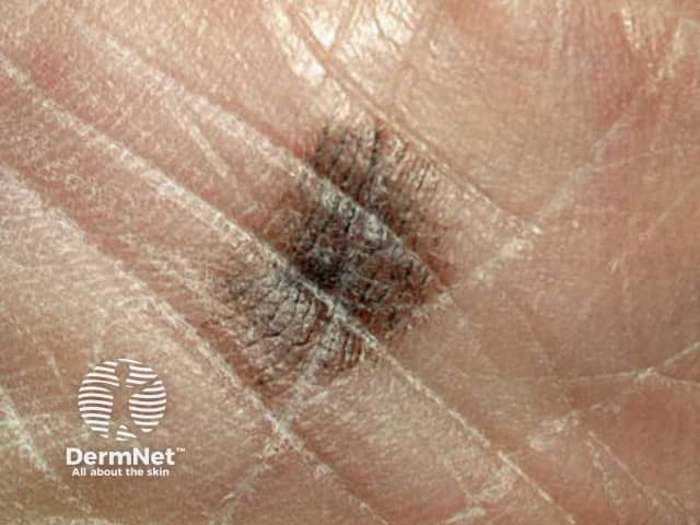
Acral lentiginous melanoma (ALM) is a rare subtype of melanoma arising on the palms, soles, or under the nails. It is also known as acral melanoma.
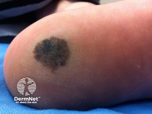
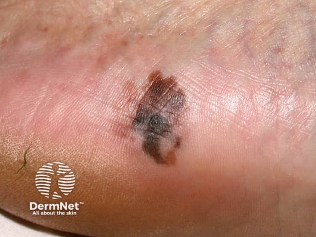
In early stages, ALM often appears as a dark brown or black macule with variegated colour, and can be nodular and ulcerated in advanced cases. ALM is slow-growing, and can take months to years to become invasive, ie, to grow beyond the basement membrane of the epidermis where it originates.
Who gets acral lentiginous melanoma?
Acral lentiginous melanoma (ALM) is the least common subtype of melanoma diagnosed overall, accounting for 2–3% of melanoma diagnoses.
However, ALM is the most common subtype of melanoma in darker skinned populations, accounting for 40–60% of melanoma diagnoses in Asian and African-American ethnicities. It is the least common subtype of melanoma in Caucasian populations.
The incidence of ALM increases with age, with the average age of diagnosis being 63 years. Males and females are equally affected. Females tend to be diagnosed earlier than males.
What causes acral lentiginous melanoma?
Acral lentiginous melanoma (ALM) usually arises de novo, but there are rare reports of acral melanoma arising from a pre-existing melanocytic naevus.
The cause of ALM is not fully understood. Unlike other subtypes of melanoma, sun exposure does not appear to be a risk factor for its development, however, there are rare case reports demonstrating an ultraviolet signature as in other types of cutaneous melanoma.
Suggested aetiologies of the cause for ALM include:
- Stress/shearing forces on the foot
- Genetic mutations, including KIT, BRAF, NRAS, NF1, cyclin D1, CDKN2A, and MYC.
The pathogenesis for melanoma includes overgrowth of melanocytes, the pigment-producing cells found deep in the epidermis.
What are the clinical features of acral lentiginous melanoma?
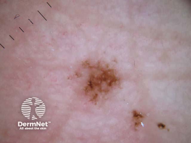
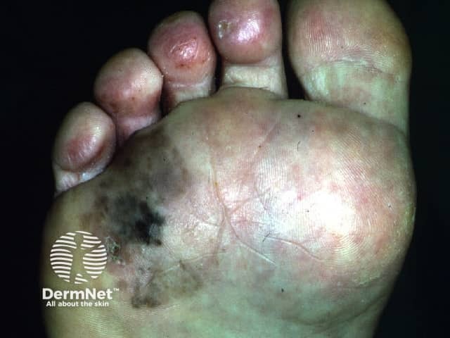
In early stages, acral lentiginous melanoma (ALM) presents as a new light to dark brown pigmented macule on the palm, sole, or under a nail that follow the skin ridges. ALM is more commonly seen on the feet, possibly due to higher melanocyte density.
Occasionally, ALM can present as an amelanotic or hypomelanotic lesion. These are difficult to diagnose as they often mimic benign diseases such as warts or calluses.
Subungual melanoma arises from the nail matrix and usually presents as a longitudinal brown or black band in the fingernail or toenail. It may be associated with nail dystrophy. Involvement of the proximal nail fold (Hutchinson’s sign) is considered a clue to the diagnosis.
Suspicious features for invasive melanoma:
- Nodular or darkly pigmented appearance
- Ulceration
- Pruritus
- Increasing number of colours, particularly blue and black
- Pigment extending from under the nail to beyond the nail fold
- >7mm in size.
How do clinical features vary in differing types of skin?
Clinical features do not vary significantly between skin types. It may be more difficult to visualise naevus structure on dermoscopy in darker skin types, appearing as a more homogenous colour.
What are the complications of acral lentiginous melanoma?
Complications of untreated disease:
- Progressive disease including distal metastasis.
Complications of surgical management:
- Scarring
- Skin contractures
- Digital amputation
- Phantom limb pain
- Functional impairment
- Loss of nail/s.
How is acral lentiginous melanoma diagnosed?
The diagnosis is often made on clinical history and presentation, supported by dermoscopy.
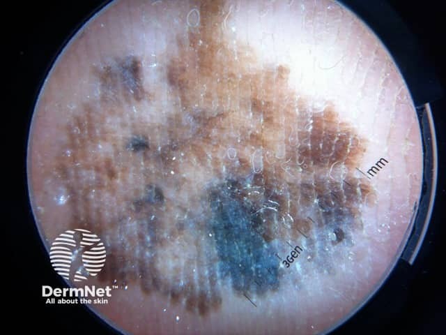
The following features are seen on dermoscopy for acral lentiginous melanoma (ALM):
- Parallel ridge pattern in early phases
- Lack of features typical of acral naevi (fibrillary, lattice-like, parallel furrow patterns)
- Blotches
- Asymmetry
- Multiple colours
- Diffuse pigmentation of the nail/stripes on the nail/nail ulceration.
Definitive diagnosis is made with histopathology after excision or biopsy of the lesion. Staging is conducted as for any other cutaneous melanoma, with histopathological features such as Breslow depth, ulceration, and sentinel lymph node status.
Histological features of ALM include an asymmetrical proliferation of melanocytes at the dermoepidermal junction.
What is the differential diagnosis for acral lentiginous melanoma?
The differential diagnosis for acral lentiginous melanoma is broad and includes the following:
- Acral naevi (either congenital or acquired)
- Trauma-related haemorrhage
- Calcaneal petechiae (talon noir)
- Tinea nigra
- Soil/stain.
Of note, pigmented actinic keratoses and seborrhoeic keratoses do not occur on acral surfaces.
Amelanocytic or hypomelanocytic lesions may mimic benign diseases such as warts, calluses, or benign tumours such as poromas.
What is the treatment for acral lentiginous melanoma?
General measures
- Education of health professionals and the public.
- Regular skin examinations.
- Multidisciplinary management with a podiatrist and occupational therapist where available.
Specific measures
The mainstay of treatment for acral lentiginous melanoma (ALM) is wide local excision. Further treatment depends mainly on the Breslow thickness of the lesion.
After initial excision biopsy, the radial excision margins, measured clinically from the edge of the melanoma, are:
- In-situ lesions: 0.5–1 cm margin
- Breslow depth <1 mm: 1 cm margin
- Breslow depth 1–2mm: 1–2 cm margin
- Breslow depth >2mm: 2 cm margin.
A skin graft or a flap may be needed to close the wound, or in some cases, partial amputation of a digit.
If further treatment is required following excision, this may involve radiotherapy or immunotherapy.
How do you prevent acral lentiginous melanoma?
While there are no known preventative strategies, regular skin checks and general sun protection are recommended. Any new lesions noted on the palms, soles, or under the nails should be reviewed by a medical professional.
What is the outcome for acral lentiginous melanoma?
Compared with cutaneous melanoma, most studies suggest that acral lentiginous melanoma has a poorer prognosis, with diagnosis often occurring at a more advanced clinical stage. It is unclear if this is solely due to delay in ALM detection due to the difficulty in monitoring such sites (eg, soles of the feet), or is also a consequence of genetic and biological differences that promote tumour aggressiveness.
The outcome of ALM depends primarily on Breslow thickness of the lesion and staging, including sentinel lymph node involvement. Other factors that may influence prognosis include age and gender.
Bibliography
- Basurto-Lozada P, Molina-Aguilar C, Castaneda-Garcia C, et al. Acral lentiginous melanoma: Basic facts, biological characteristics and research perspectives of an understudied disease. Pigment Cell Melanoma Res. 2021;34(1):59–71. doi: 10.1111/pcmr.12885. Journal
- Bernardes SS, Ferreira I, Elder DE, et al. More than just acral melanoma: the controversies of defining the disease. J Pathol Clin Res. 2021;7(6):531–541. doi: 10.1002/cjp2.233. Journal
- Hall KH, Rapini RP. Acral Lentiginous Melanoma. In: StatPearls. Treasure Island (FL): StatPearls Publishing; 2022. Available here
- Kolla AM, Vitiello GA, Friedman EB, et al. Acral Lentiginous Melanoma: A United States Multi-Center Substage Survival Analysis. Cancer Control. 2021;28:10732748211053567. doi: 10.1177/10732748211053567. Journal
- Lobl MB, Santos C, Clarey D, et al. Treatments and associated outcomes of acral lentiginous melanoma: A review. J Am Acad Dermatol. 2020;83(1):230–234. doi: 10.1016/j.jaad.2019.11.021. Journal
- Newell F, Wilmott JS, Johansson PA, et al. Whole-genome sequencing of acral melanoma reveals genomic complexity and diversity. Nat Commun. 2020;11(1):5259. doi: 10.1038/s41467-020-18988-3. Journal
- Tas F, Erturk K. Acral Lentiginous Melanoma Is Associated with Certain Poor Prognostic Histopathological Factors but May Not be Correlated with Nodal Involvement, Recurrence, and a Worse Survival. Pathobiology. 2018;85(4):227–231. doi: 10.1159/000488457. Journal
On DermNet
- Acral lentiginous melanoma images
- Acral lentiginous melanoma dermoscopy
- Skin cancer
- Melanoma
- Melanoma pathology
- Melanoma in situ
- Melanoma of the nail unit
- Melanoma in skin of colour
- Genetics of melanoma
- Other melanoma resources
Other websites
- Melanoma risk assessment tool — Melanoma Institute of Australia
- Melanoma education portal — Melanoma Institute of Australia
- Acral lentiginous melanoma — Memorial Sloan Kettering Cancer Center
- Clinical Guidelines — Cancer Council Australia