Introduction
Demographics
Causes
Clinical features
Variation in skin types
Complications
Diagnosis
Differential diagnoses
Treatment
Outcome
Retiform purpura is a cutaneous sign that falls within the spectrum of reticulate eruptions of vascular origin.
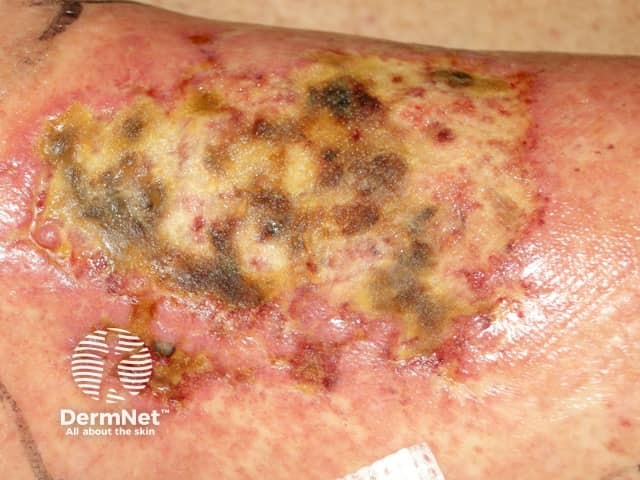
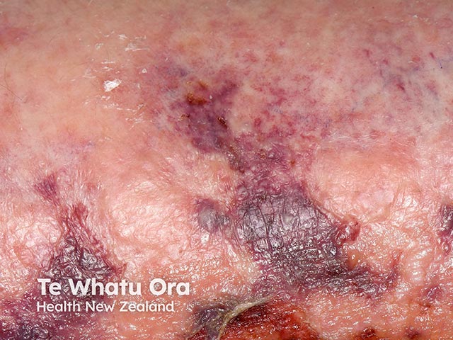
It comprises non-blanching, purpuric patches or plaques that are angulated or branching (reticular), often accompanied by skin necrosis and ulceration. Retiform purpura occurs when cutaneous blood vessels are compromised, resulting in downstream skin ischaemia.
The demographic group affected depends on the underlying diagnosis. Adults and children can present with retiform purpura.
Retiform purpura results from either blood vessel wall damage or blood vessel lumen occlusion, causing vessel compromise and downstream skin ischaemia, purpura, and/or necrosis.
Retiform purpura has been reported in seriously ill COVID-19 patients due to microthrombi formation.

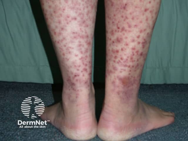
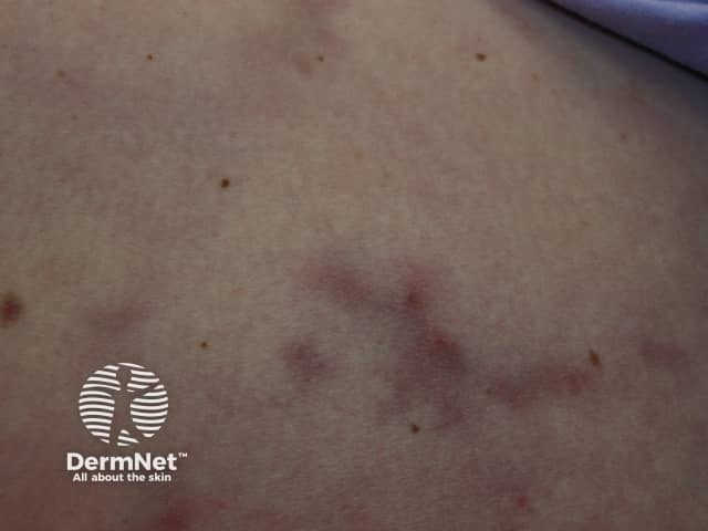
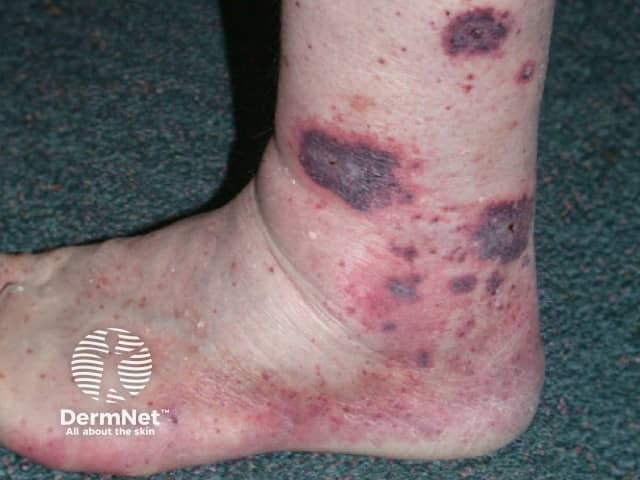
Retiform purpura presents as painful dark red or purple patches or plaques, which can vary in size from small (1–2 cm) to large (>10 cm), may be single or multiple, and may be localised or disseminated.
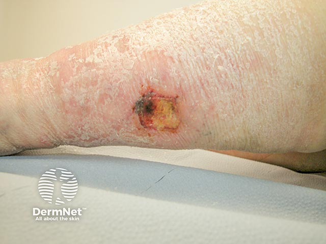
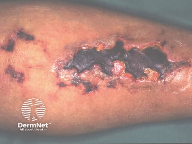
Retiform purpura displays branching or angulation at the periphery with a purpuric center, often accompanied by frank or impending ulceration and/or necrosis. This characteristic morphology is an important clue for diagnosing retiform purpura and any potentially serious underlying condition.
The acute, widespread development of retiform purpura (termed ‘purpura fulminans’) represents a rare, life-threatening emergency. Purpura fulminans is a severe and rapidly fatal form of disseminated intravascular coagulation (DIC) with pronounced skin necrosis, sometimes associated with symmetrical peripheral gangrene.
Clinical clues to the underlying cause may include the following:
Patient factors
Stage of lesions
Lesion morphology
Lesion distribution
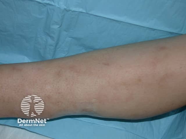
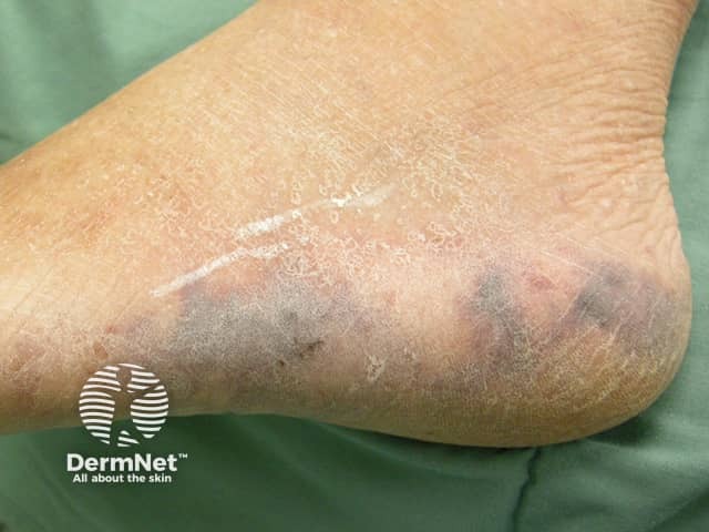
Retiform purpura is not well described in darker skin types. However, case reports have noted that the purpura presents as a darker purple colour rather than the bright red that is often observed in lighter skin types.
Retiform purpura can be complicated by:
Additional complications will depend on the underlying cause.
Retiform purpura is a clinical diagnosis based on typical appearance. Investigations are required to determine the underlying cause and will be guided by the following:
Routine tests may include:
A skin biopsy taken from the peripheral purpuric rim should be performed; the histology varies with the underlying diagnosis. Examples include warfarin necrosis pathology, calciphylaxis pathology, and cholesterol emboli pathology.
The differential diagnosis of retiform purpura includes other entities within the spectrum of reticulate eruptions as well as non-reticulate morphologies that share similar underlying ischaemic features:
Ulcerated skin lesions may require appropriate wound dressings and debridement of necrotic tissue. Circulatory support and blood product replacement may be necessary.
Treatment of retiform purpura is aimed at treatment of the underlying cause.
Outcomes depend on the underlying cause of the retiform purpura and can range from full recovery (eg, removing precipitants such as emboli secondary to a procedure) to death, as may occur in a catastrophic disseminated opportunistic infection.

