DermNet provides Google Translate, a free machine translation service. Note that this may not provide an exact translation in all languages

Translate

Epidermolysis bullosa acquisita

Author: Vanessa Ngan, Staff Writer, 2003.


toc-icon

What is epidermolysis bullosa?

Epidermolysis bullosa (EB) is the name given to a group of inherited blistering diseases that are present from birth. 

What is epidermolysis bullosa acquisita?

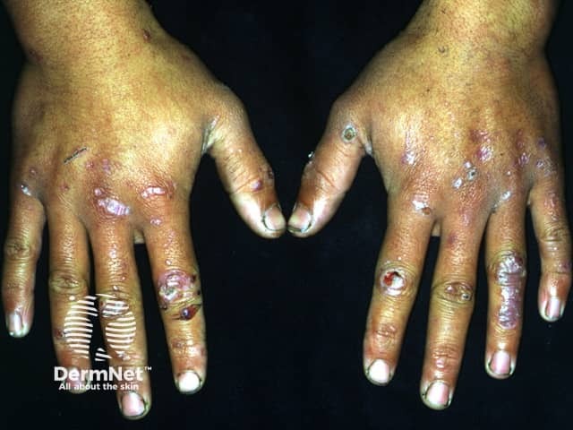
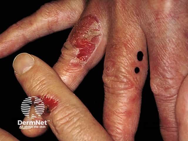
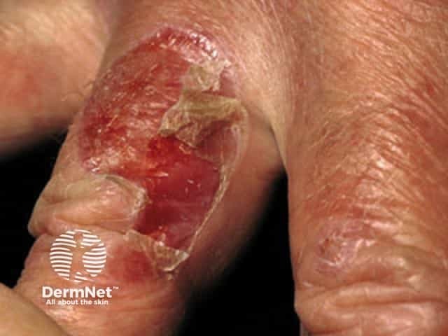
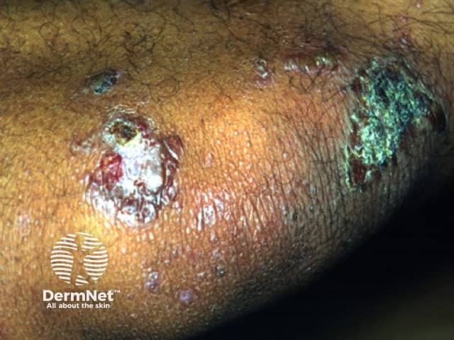
Epidermolysis bullosa acquisita (EBA) is a rare autoimmune blistering disease in which tense subepithelial blisters appear at sites of traumaUnlike EB, EBA is not inherited and usually presents in adult life.

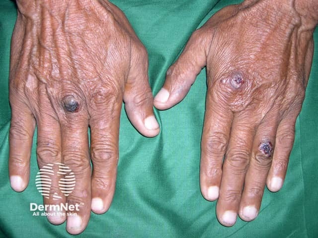
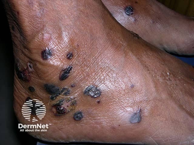
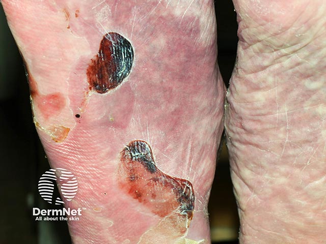
EBA blisters tend to be localised to areas that are easily injured such as the hands, feet, knees, elbows, and buttocks. Sometimes there is mucosal involvement with blisters forming in the mouth, nose and eyes. 

Epidermolysis bullosa acquisita

Who gets epidermolysis bullosa acquisita and why?

EBA usually occurs in the fourth and fifth decade of life. Males and females of all races can be affected.

Some patients with EBA have been reported to have other autoimmune diseases, most often Crohn disease and systemic lupus erythematosus, or health problems such as amyloidosis, multiple myeloma and, rarely, carcinoma of the lung and lymphoma. Other patients only have a skin problem. 

What is the cause of epidermolysis bullosa acquisita?

Researchers have detected tissue-bound immunoglobulin (Ig)-G autoantibodies directed against the NC1 domain of type VII collagen (Col7) in affected patients. Type VII collagen is the major structural component of anchoring fibrils. These attach the basement membrane of the epidermis to the underlying dermis. The loss of anchoring fibrils leads to the formation of blisters just under the epidermis within an area known as the lamina densa

What are the clinical features of epidermolysis bullosa acquisita?

EBA has several distinct clinical presentations.

Classification of EBA Clinical features

Classical mechanobullous form of EBA

  • This is the most common form of EBA in Europeans
  • Tense vesicles and bullae primarily on extensor surfaces of hands, knees, knuckles, elbows and ankles
  • Mucous membrane blisters rupture easily
  • Lesions heal with significant scarring and milia (small white spots)
  • Resembles the inherited form of dystrophic epidermolysis bullosa

Non-classical/non-mechanobullous form of EBA

  • Widespread tense vesicles and bullae not localised to trauma-prone sites
  • Often affects the trunk and skin folds
  • Generalised redness, itching and plaque formation
  • Heals with minimal scarring and milia formation
  • Resembles bullous pemphigoid

Predominantly mucous membrane form of EBA

  • Mucous membranes affected include buccal, gingival, palatal, conjunctival, nasopharyngeal, oesophageal, anus and genital
  • May result in significant mucosal scarring and dysfunction
  • Resembles mucous membrane pemphigoid

Brunsting‐Perry‐type EBA

  • Chronic recurrent blistering disease
  • Confined to head and neck
  • Resembles Brunsting-Perry type mucous membrane pemphigoid

IgA‐EBA

  • Resembles linear IgA bullous dermatosis
  • Linear IgA deposits are found in the basement membrane zone
  • Tends to be aggressive with mucosal scarring

How is epidermolysis bullosa acquisita diagnosed?

The following tests should be performed to establish a diagnosis of EBA.

  • Skin biopsy — this shows the presence of a blister under the epidermis (subepidermal bulla); a variable infiltrate of mixed inflammatory cells in the dermis; and milia cysts and scarring in older lesions.
  • Direct immunofluorescence (DIF) of a skin biopsy — this shows deposition of immunoglobulin (IgG more often than IgM or IgA) and C3  in a line along the dermoepidermal junction. Blood vessels are uninvolved.
  • Indirect immunofluorescence (IIF) of serum — this reveals low titre circulating IgG autoantibodies that target the basement membrane.

Other tests to confirm the diagnosis are only available in some academic centres. They include [2]:

  • Skin biopsy for direct and indirect immunoelectron microscopy, serration pattern analysis by DIF,  fluoroescent overlay antigen mapping, and DIF on salt-split skin
  • Blood tests to detect circulating antibodies to Col7: immunoblotting, enzyme-linked immunosorbent assay (ELISA), more detailed IIF, and immunoprecipitation.

The International Bullous Diseases Group (IBDG) have proposed by consensus clinical and diagnostic criteria for EBA [1,2]. If a blistering disease is clinically and histologically compatible with EBA, the diagnosis is confirmed if: 

  • Linear IgG and/or C3 deposition along the basement membrane zone is demonstrated by direct immunofluorescence (DIF), and,
  • Autoantibodies against COL7 are detected in the patient's serum (via enzyme‐linked immunosorbent assay).

What is the differential diagnosis for epidermolysis bullosa acquisita?

EBA may mimic other inflammatory blistering diseases, most often bullous pemphigoid and bullous systemic lupus erythematosus (which is also associated with anti-Col7 autoantibodies).

In comparison to bullous pemphigoid, EBA [2]:

  • Arises in younger individuals, usually under the age of 70 years
  • Has prominent involvement of the head and neck
  • Involves mucous membranes
  • Lesions heal with scarring and the formation of milia.

What is the treatment for epidermolysis bullosa acquisita?

The primary aim in the treatment of EBA is to protect the skin and stop blister formation, promote healing and prevent complications.

Because EBA is considered an autoimmune disease it is reasonable to use immunosuppressive agents to modify or reduce autoimmune responses and decrease the production of autoantibodies. These may include:

Due to the rarity of the disease, it is difficult to be sure which drug is the most effective.

Other important management strategies include:

  • Treatment of any underlying disease.
  • Consultation with other specialists such as dentist and ophthalmologist.
  • Avoid direct physical trauma to skin surfaces.
  • Avoid hard or brittle food or food with high acid content in patients with oral involvement.

What is the prognosis?

EBA is a chronic inflammatory disease that has periods of partial remissions and exacerbations. If treated and cared for properly, patients can expect to live a normal lifespan. Problems that may arise include:

  • Infection of open wounds and sores
  • Severe scarring which may cause deformities and restriction of movement
  • Periodontal disease
  • Conjunctival scarring and blindness
  • Side effects of medication


References

  1. Yamagami J. The International Bullous Diseases Group consensus on diagnostic criteria for epidermolysis bullosa acquisita: a useful tool for dermatologists. Br J Dermatol. 2018 Jul;179(1):7. doi: 10.1111/bjd.16440. PubMed PMID: 30156286.
  2. Prost-Squarcioni C, Caux F, Schmidt E et al. International Bullous Diseases Group: consensus on diagnostic criteria for epidermolysis bullosa acquisita. Br J Dermatol. 2018 Jul;179(1):30-41. doi: 10.1111/bjd.16138. Epub 2018 May 8. Review. PubMed PMID: 29165796.
  3. Textbook of Dermatology. Ed Rook A, Wilkinson DS, Ebling FJB, Champion RH, Burton JL. Fourth edition. Blackwell Scientific Publications.

On DermNet

Other websites


Books about skin diseases


Related information

Sign up to the newsletter