Main menu
Common skin conditions

NEWS
Join DermNet PRO
Read more
Quick links
Author(s): Dr Prudence Gramp, Dermatology Department, Gold Coast University Hospital, Australia. Copy edited by Gus Mitchell. May 2022
Introduction
Demographics
Causes
Clinical features
Variation in skin types
Complications
Diagnosis
Differential diagnoses
Treatment
Outcome
Polymorphic light eruption (PMLE) is a seasonal, acquired, idiopathic photodermatosis occurring in spring and early summer.
It is also known as polymorphous light eruption, sun allergy, sun poisoning, prurigo aestivalis, summer eruption/prurigo, or eczema solare. Juvenile spring eruption is a variant of PMLE.

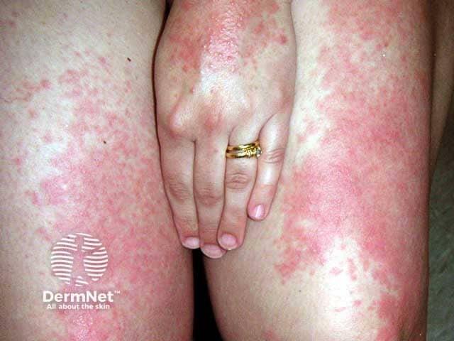
Extensive papular and plaque polymorphic light eruption

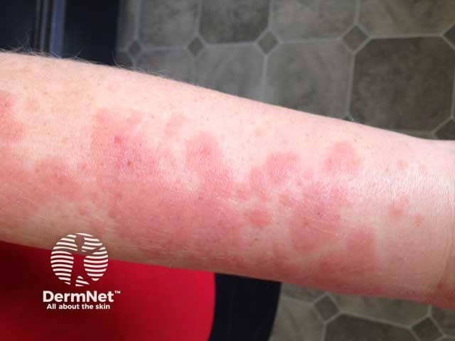
Targetoid polymorphic light eruption on recently exposed arm skin
Patients with PMLE can develop a tolerance during summer months.
There is a genetic susceptibility in 15–46% of cases where a positive family history is reported.
PMLE is a delayed hypersensitivity reaction in the skin to unknown endogenous cutaneous photo-induced antigens. This abnormal response to ultraviolet (UV) light means affected patients develop an inflammatory response to an endogenous photo-induced antigen. The following factors must be considered when determining pathogenesis and when implementing protective measures:
UV radiation usually creates an immunosuppressive response in the skin, however, patients with PMLE may have a reduction in this normal response. In PMLE patients, UV radiation leads to an increased amount of CD4 and CD8 T lymphocytes, and an increased inflammatory response in the epidermis and dermis. The photo antigen that triggers this response is currently unknown.
Some patients experience PMLE during phototherapy, which is used to treat skin conditions such as psoriasis and dermatitis.
Because PMLE is more prevalent in women than men, it is hypothesized that there is a hormonal component to its pathogenesis. Estradiol may act as an inhibitor to the UV light immunosuppression which would normally aid in reducing hypersensitivity reactions.
As the name suggests, clinical features can vary — poly meaning “many”, morphic meaning “forms”. The morphology can include eruptions that are:
The morphology is, however, always the same in one patient.
Distribution can include areas exposed to sunlight such as the arms, lower legs, V of the neck, and the chest. The dorsal hands and face are uncommon sites for PMLE possibly due to their chronic exposure to the sun and hardening of the skin. The eruption is usually symmetrically distributed in a patchy fashion and typically does not involve all of the exposed skin. It can be mildly to markedly pruritic and general malaise, headache, fever, and nausea can occur in rare cases.
PMLE persists for several days and can worsen if the affected skin is exposed to further sunlight before resolution of the previous eruption. It resolves without scarring.
There is a phenomenon called the skin hardening effect where chronic exposure to sunlight leads to skin changes including increased melanin and thickening of the stratum corneum. These changes are thought to restore the skin’s normal immunosuppressive response to UV light and hence reducing or resolving PMLE over time. This can explain why it is uncommon to get PMLE in areas of the face or hands due to their chronic exposure to the sun compared to other areas of the body.
PMLE can be seen in all races and all skin types. It is more common in people with lighter skin. In darker skin types, the most common morphology is grouped, pinhead-sized papules.
A clinical diagnosis of polymorphic light eruption can be made based on a history of a pruritic eruption occurring following sun exposure and previous episodes in spring or summer. Accurate diagnosis relies on the exclusion of other photosensitive conditions.
To exclude other photosensitive conditions a skin biopsy may be considered. The histopathology of PMLE is nonspecific, variable, and can include:
Direct immunofluorescence is negative in PMLE.
Suitable investigations to determine the exclusion of cutaneous lupus erythematosus include full blood count; circulating antinuclear antibodies (ANA); extractable nuclear antigens (ENA); and direct immunofluorescence on histopathology.
Phototesting can be considered but is not carried out in all patients with PMLE.
PMLE may be lifelong although 60% of people see improvement or resolution over 15 years and 75% of people in 30 years.
The eruption can appear within hours of sun exposure and last for days. It can worsen with repeated exposure to sunlight before the eruption has resolved. It has been noted that PMLE appears to be less frequent and severe in women after menopause.