DermNet provides Google Translate, a free machine translation service. Note that this may not provide an exact translation in all languages

Translate

Reactive perforating collagenosis

Authors: Vanessa Ngan, Staff Writer, 2005; Updated: Dr Aswan Tai, Junior Doctor, Peter MacCallum Cancer Centre, Melbourne, Australia; Dr Sarah Smithson, Dermatologist, Skin Health Institute, Melbourne, Australia. Copy edited by Gus Mitchell. September 2021


toc-icon

What is reactive perforating collagenosis?

Reactive perforating collagenosis is the most common type of primary perforating dermatosis. It is characterised by the transepithelial elimination of collagen from the dermis through the epidermis to the skin surface.

Reactive perforating collagenosis

Who gets reactive perforating collagenosis?

  • Familial reactive perforating collagenosis
    • Rare
    • Autosomal dominant or autosomal recessive inheritance and sporadic cases
    • Onset is usually in infancy or early childhood
  • Acquired reactive perforating collagenosis
    • More common form
    • Onset is usually in adulthood
    • Female predominance 3:1
    • No clear racial predilection
    • Strong association with diabetes mellitus and chronic renal disease; 10% of haemodialysis patients develop reactive perforating collagenosis
    • Other reported associations include concurrent itchy dermatoses, malignancies, pregnancy and endocrine disorders, infections and infestations, and medications

What causes reactive perforating collagenosis?

The pathogenesis of reactive perforating collagenosis is unclear.

The inherited form appears to be due to a genetic abnormality in collagen causing focal damage and extrusion through the epidermis. Cold weather and skin trauma typically trigger or aggravate the skin lesions.

Microvascular insufficiency and elevated fibronectin levels in plasma, as seen with diabetes and renal failure, may play a role in the acquired form. Another theory suggests microdeposition of substances and abnormal glycosylation of collagen I and III in diabetes alter collagen fibres.

Superficial trauma, such as scratching, and cold leads to necrobiosis and epidermal thinning in susceptible patients.

What are the clinical features of reactive perforating collagenosis?

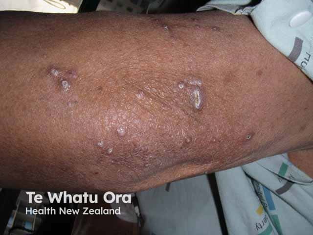
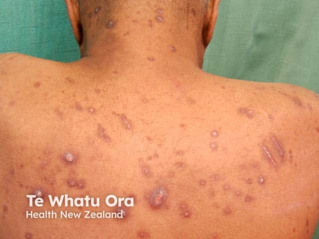
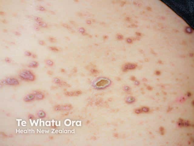
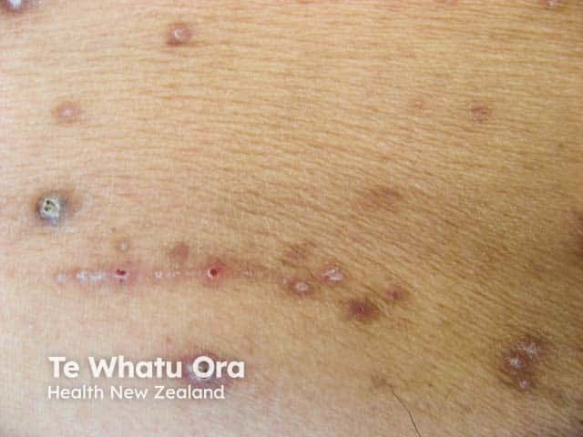
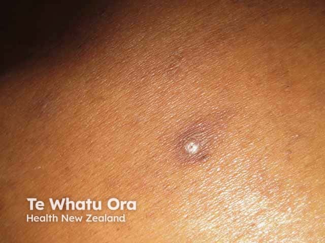
Reactive perforating collagenosis is a papulonodular mucocutaneous disorder with adherent keratotic plugs and crusts.

  • Red-brown umbilicated papules and nodules, often with a cup-shaped depression, central keratotic crust, and erythematous halo
  • Variable in shape and size, usually up to 10 mm in diameter; a rare giant form has been described with individual lesions up to 10 cm in diameter
  • Lesions are typically intensely itchy; pain or tenderness is rare
  • Koebner phenomenon is a common feature
  • Commonly located on trunk or extensor aspect of the limbs but can be widespread occurring anywhere on the skin
  • Hands, elbows, and knees are the common initial sites involved in the inherited form
  • Mucosal lesions have been described
  • Variable phenotype in the familial form, even within the one family

Clinical features of reactive perforating collagenosis

Dermoscopy features of reactive perforating collagenosis

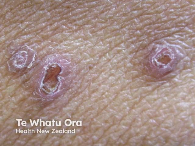
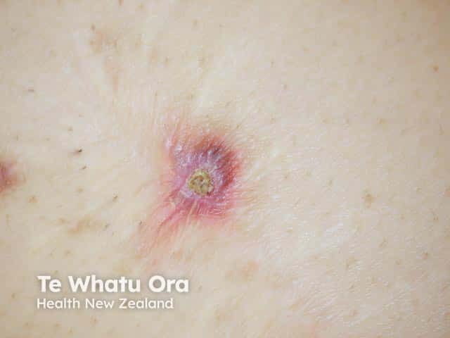
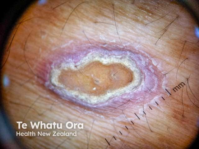
The dermoscopy features of reactive perforating collagenosis are characteristic and consistent:

  • Yellow-brown structureless area in the centre of the lesion matching the central crust
  • White rim of variable thickness around the central crust indicating keratinous debris or epidermal invagination
  • Outer pink circle with short looped and peripheral dotted vessels corresponding with dermal inflammation.

Dermoscopy of reactive perforating collagenosis

How do clinical features vary in differing types of skin?

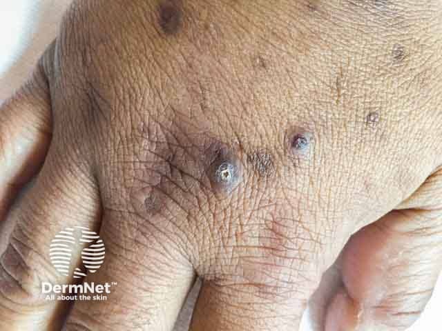
Reactive perforating collagenosis in skin of colour is associated with hyperpigmentation:

  • Of lesions secondary to scratching
  • Of scratch marks
  • Following resolution of lesions.

Hyperpigmentation related to reactive perforating collagenosis in skin of colour

What are the complications of reactive perforating collagenosis?

  • Impact of chronic itch on quality of life
  • Secondary bacterial skin infection
  • Healing of individual lesions with scarring and/or dyspigmentation in all skin types

How is reactive perforating collagenosis diagnosed?

Reactive perforating collagenosis can usually be diagnosed on its distinct clinical features and associated conditions, and confirmed on dermoscopy.

Multiple skin biopsies examined with multiple levels may be required to find the diagnostic histology [see Reactive perforating collagenosis pathology].

What is the differential diagnosis for reactive perforating collagenosis?

What is the treatment for reactive perforating collagenosis?

The main aim of treatment for reactive perforating collagenosis is to reduce itch and minimise skin trauma:

Treatment of individual lesions

Preventive measures

  • Treatment of the associated systemic condition

What is the outcome for reactive perforating collagenosis?

Reactive perforating collagenosis remains confined to the skin.

Familial reactive perforating collagenosis is a lifelong condition with lesions becoming larger and more numerous with age.

Individual lesions in both the familial and acquired forms are self-healing but often recur.


Bibliography

  • Bhat YJ, Manzoor S, Qayoom S, Wani R, Baba AN, Bhat AH. Familial reactive perforating collagenosis. Indian J Dermatol. 2009;54(4):334–7. doi:10.4103/0019-5154.57608. Journal
  • Karpouzis A, Giatromanolaki A, Sivridis E, Kouskoukis C. Acquired reactive perforating collagenosis: current status. J Dermatol. 2010;37(7):585–92. doi:10.1111/j.1346-8138.2010.00918.x. PubMed 
  • Lukács J, Schliemann S, Elsner P. Treatment of acquired reactive perforating dermatosis - a systematic review. J Dtsch Dermatol Ges. 2018;16(7):825–42. doi:10.1111/ddg.13561. PubMed
  • Ormerod E, Atwan A, Intzedy L, Stone N. Dermoscopy features of acquired reactive perforating collagenosis: a case series. Dermatol Pract Concept. 2018;8(4):303–5. doi:10.5826/dpc.0804a11. PubMed Central
  • Ramesh V, Sood N, Kubba A, Singh B, Makkar R. Familial reactive perforating collagenosis: a clinical, histopathological study of 10 cases. J Eur Acad Dermatol Venereol. 2007;21(6):766–70. doi:10.1111/j.1468-3083.2006.02085.x. PubMed
  • Rapini RP, Herbert AA, Drucker CR. Acquired perforating dermatosis. Evidence for combined transepidermal elimination of both collagen and elastic fibers. Arch Dermatol. 1989;125(8):1074–8. doi:10.1001/archderm.125.8.1074. PubMed
  • Satchell AC, Crotty K, Lee S. Reactive perforating collagenosis: a condition that may be underdiagnosed. Australas J Dermatol. 2001;42(4):284–7. doi:10.1046/j.1440-0960.2001.00537.x. PubMed

On DermNet

Other websites

Books about skin diseases


Related information

Sign up to the newsletter