

ABCDEFG of melanoma — extra information
Introduction Other skin lesions with ABCDEFG features Melanomas without ABCDEFG features Importance What to do about concerning lesions
What is the 'ABCDEFG' of melanoma?
The 'ABCDE' of melanoma is an acronym designed to help the public and clinicians identify features in a skin lesion that may suggest an early or in situ melanoma (superficial spreading melanoma, lentigo maligna melanoma, or acral lentiginous melanoma).
- Asymmetry
- Border irregularity
- Colour variability / Change
- Different
- Evolving
Melanoma ABCD signs

The EFG of melanoma is another acronym designed to help the public and clinicians identify skin changes in a lesion suggestive of nodular melanoma.
- Elevated
- Firm
- Growing
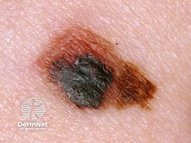
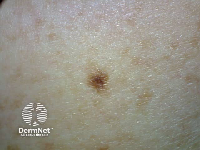
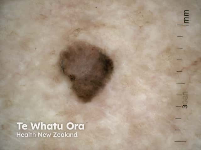
Asymmetry
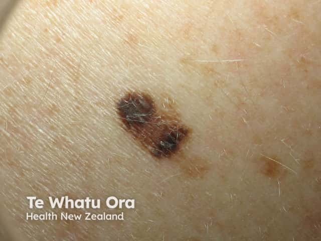
A is for Asymmetry.
A melanocytic naevus (harmless mole) is usually symmetrical, whereas melanoma is often irregular or asymmetrical in shape and/or colour.
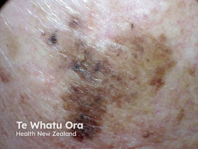
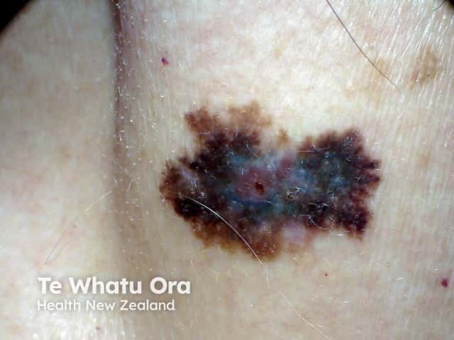
Asymmetry of shape and/or colour in a melanoma
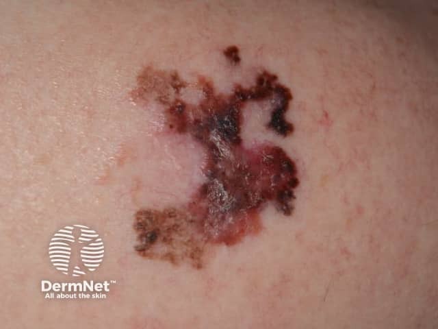
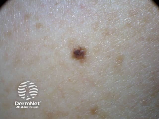
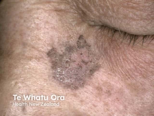
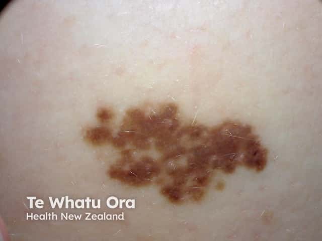
Border irregularity
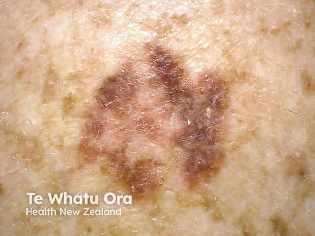
B is for Border irregularity.
A melanocytic naevus has smooth, even borders, whereas a melanoma often has irregular, blurry, or jagged edges and hard-to-define border.
On careful inspection, the pigmented component of a flat melanocytic naevus fades out towards the edge, whereas the edges of a solar lentigo or a seborrhoeic keratosis are well defined. The edges of a melanoma tend to have both well-defined and fading segments.
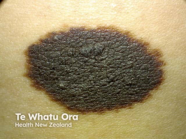
B can also used for 'black', which is an uncommon colour for a melanocytic naevus in a white-skinned individual, but may be typical in skin of colour. The colour black however can occur in seborrhoeic keratoses in all skin types and ink spot lentigo in fair skin.
Borders of pigmented skin lesions
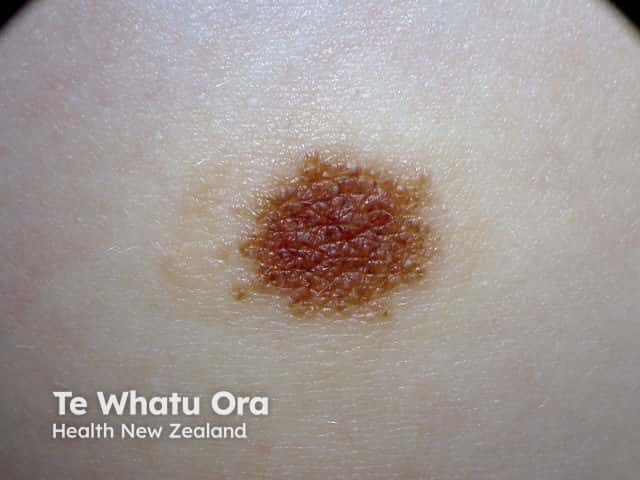
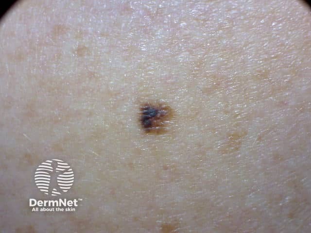
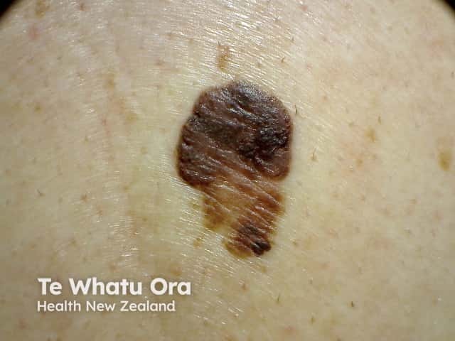
Colour variability and Changing colour
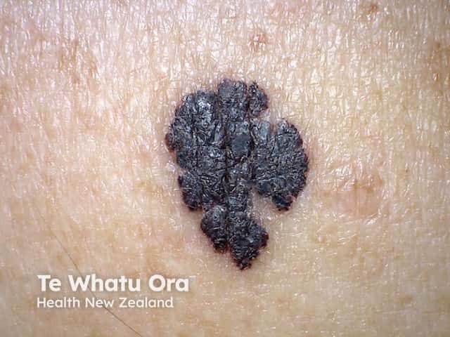
C is for Colour variability.
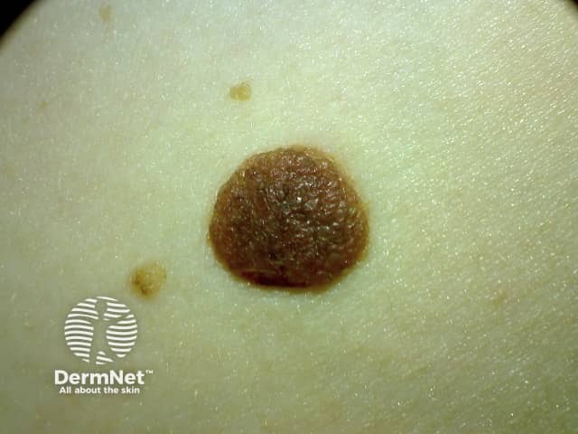
A melanocytic naevus usually has a single shade of colour or two colours with one occurring inside the other or regularly repeated (generally pink, brown, or tan).
Variation in colour of melanocytic naevi
Melanoma can be brown (96%) but can have as many as five or six colours such as blue, black, tan, grey, pink, and red: 50% of melanomas include these uncommon colours. These colours are unevenly or irregularly distributed. C is also for Changing Colour.
Variation in colour of melanoma

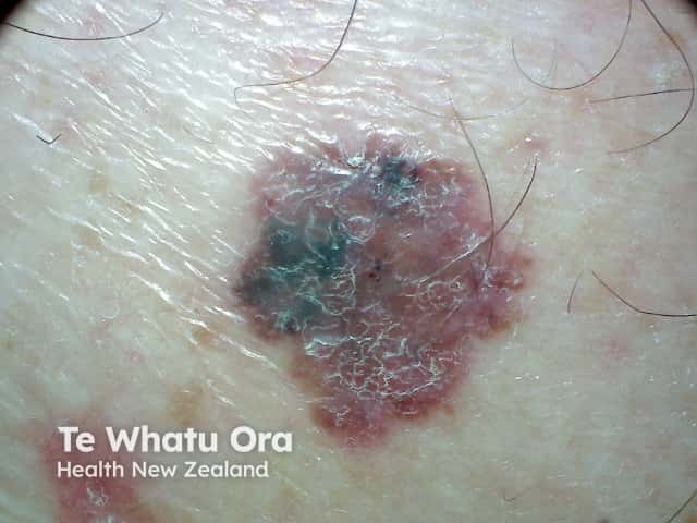
Different
D is for Different.
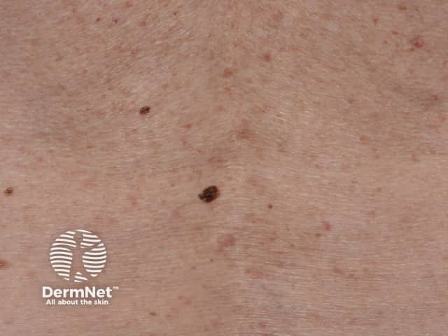
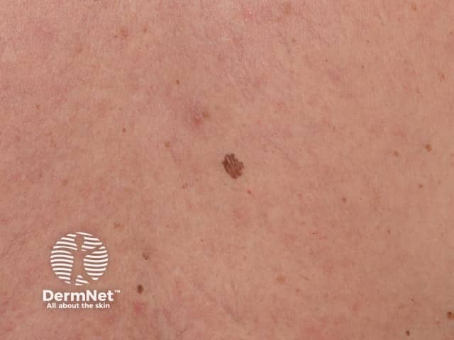
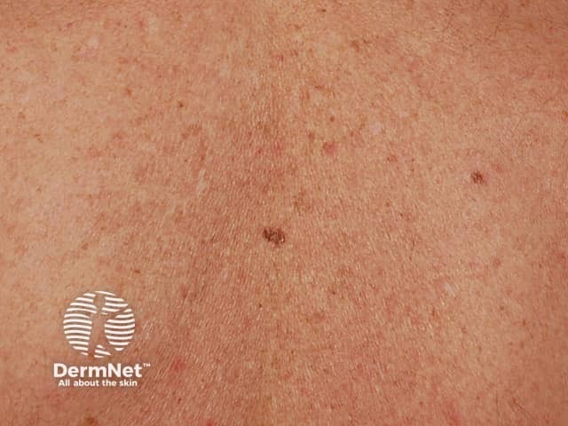
Most people have a 'signature naevus' - all their melanocytic naevi resemble each other. A melanoma appears unique and very different from the patient’s other lesions.
A pigmented lesion that is obviously different from the others is sometimes called an 'ugly duckling', 'black sheep', 'lone ranger', or 'odd-mole-out' and must be considered suspicious even if it does not fulfil the ABCDEFG criteria.
The melanoma is different from the other pigmented lesions
Evolving
E is for Evolving (changing).
A melanocytic naevus is usually stable and does not change in size, shape, or colour, whereas a melanoma changes over time. Change in size, colour, shape, or structure may be noted over months to years. However melanoma accounts for less than 3% of all changing skin lesions.
An evolving melanoma
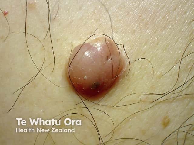
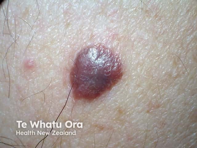
Elevated
E is for Elevated in the EFG acronym.
Benign lesions can be elevated (eg, a dermal naevus, dermatofibroma, or cyst), but a new elevated or thickened lesion may be suspicious for nodular melanoma or another form of skin cancer.
Firm
F is for Firm.
Benign lesions can feel firm (especially dermatofibroma), but this is also a feature of nodular melanoma.
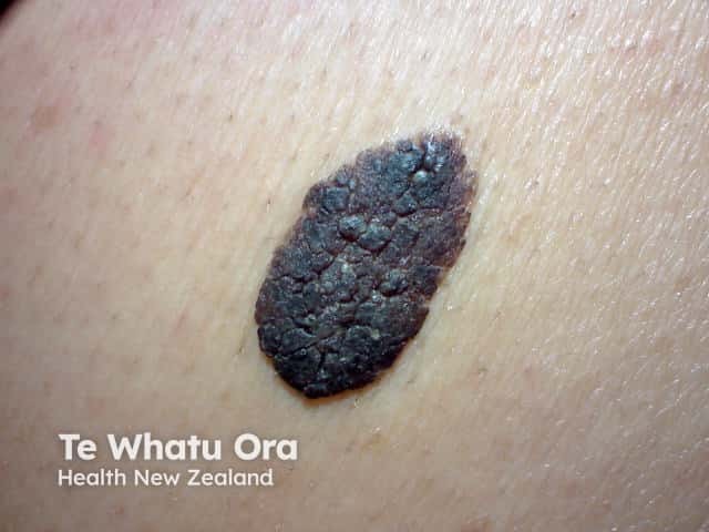
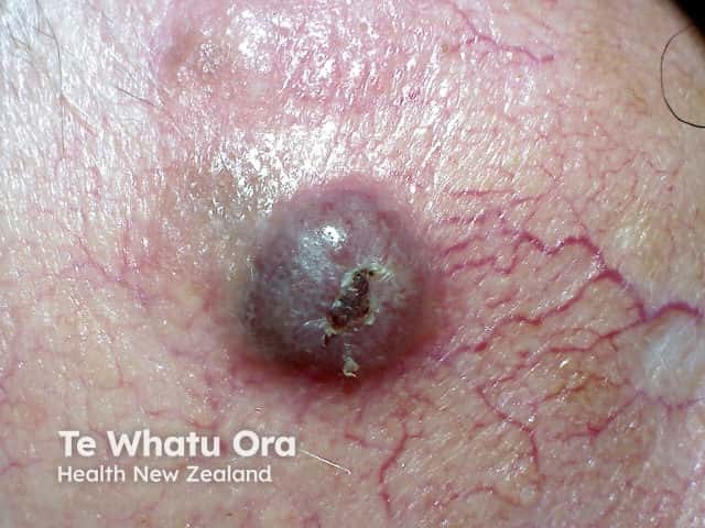
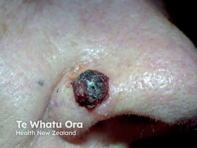
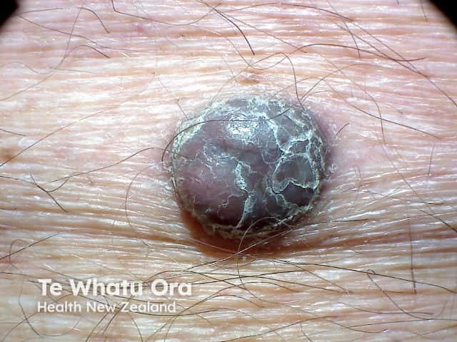
Growing
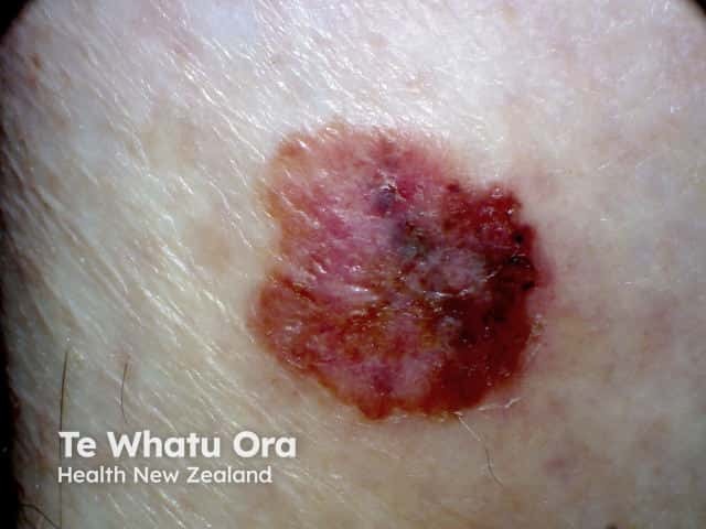
G is for Growing
A nodular melanoma tends to grow rapidly with changes noted over days or weeks. Benign skin lesions tend to remain stable or change slowly over years to decades. Although they can rapidly change in appearance over hours to days if injured, inflamed, bleeding, or affected by eczema (eg, Meyerson naevus), they do not usually grow in size.
Nodular melanoma with EFG characteristics
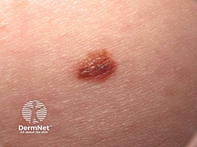
What other skin lesions might have ABCDEFG features?
The ABCDEFG criteria are not very specific for melanoma, as some or all of the criteria may be displayed by another skin cancer, such as a pigmented basal cell carcinoma or pigmented squamous cell carcinoma.
Malignant melanoma mimics
A benign lesion can be asymmetrical in shape, have an irregular border, colour variation, and be different. Examples include congenital melanocytic naevus, atypical melanocytic naevus, solar lentigo, or seborrhoeic keratosis.
Melanocytic naevi can also evolve in some circumstances such as darkening after exposure to the sun, during pregnancy, and become more elevated with age; seborrhoeic keratoses and solar lentigines routinely evolve over time.
Clinicians trained in dermoscopy can often correctly diagnose skin lesions, but sometimes a biopsy will be needed to confirm a diagnosis.
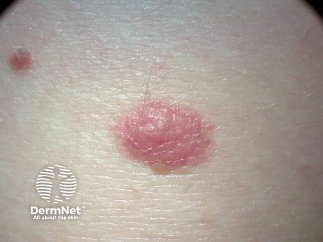
Benign melanoma mimics
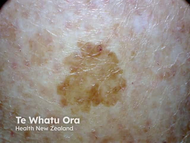
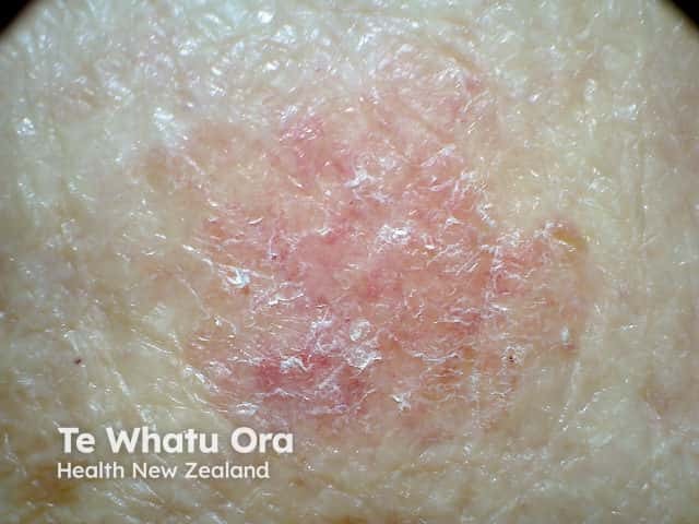
Do all melanomas display ABCDEFG characteristics?
While the ABCDEFG criteria has been proven to be very helpful in identifying a potential melanoma, they cannot be used to reliably recognise all melanomas. A melanoma may be symmetrical in shape, with a uniform border, and without much colour variation.
The ABCDEFG criteria are particularly unhelpful in the diagnosis of some less common subtypes of melanomas such as desmoplastic melanoma and melanoma in childhood, as these often lack the ABCDEFG features.
Melanomas without ABCDs
Why are these ABCDE and EFG signs important?
Melanoma is a serious form of skin cancer and can progress quickly. It is treatable if identified early but if it is untreated, it may spread to other parts of the body (metastatic melanoma) and this can be life-threatening.
Knowing the ABCDE and EFG features of melanoma can help you look for early signs of melanoma when performing a regular self-skin examination.
See our video on how to perform a self-skin examination.
I have a skin lesion I am concerned about, what should I do?
If you have a skin lesion with ABCDE or EFG characteristics that concerns you, see your doctor as soon as possible for assessment. You may be referred to a specialist for another opinion and surgery.
References
- Chamberlain AJ, Fritschi L, Kelly JW. Nodular melanoma: patients' perceptions of presenting features and implications for earlier detection. J Am Acad Dermatol. 2003;48:694–701. doi: 10.1067/mjd.2003.216. PubMed
- Gachon J, Beaulieu P, Sei JF, et al. First prospective study of the recognition process of melanoma in dermatological practice. Arch Dermatol. 2005;141:434–8. PubMed
- Grobb JJ, Bonerandi JJ. The 'ugly duckling' sign: identification of the common characteristics of nevi in an individual as a basis for melanoma screening. Arch Dermatol. 1998;134:103–4. doi: 10.1001/archderm.134.1.103-a. PubMed
- Harrington E, Clyne B, Wesseling N, et al. Diagnosing malignant melanoma in ambulatory care: a systematic review of clinical prediction rules. BMJ Open. 2017;7:e014096. doi: 10.1136/bmjopen-2016-014096. PubMed
- Jensen JD, Elewski BE. The ABCDEF rule: Combining the “ABCDE Rule” and the “ugly duckling sign” in an effort to improve patient self-screening examinations. J Clinical Aesthet Dermatol. 2015;8:15. PubMed Central
- Whited JD, Grichnik JM. The rational clinical examination. Does this patient have a mole or a melanoma? JAMA. 1998;279:696–701. doi: 10.1001/jama.279.9.696. PubMed
- Yagerman SE, Chen L, Jaimes N, Dusza SW, Halpern AC, Marghoob A. 'Do UC the melanoma?' Recognising the importance of different lesions displaying unevenness or having a history of change for early melanoma detection. Australas J Dermatol. 2014;55(2):119-24. doi:10.1111/ajd.12143 PubMed
On DermNet
- About melanoma: for patients
- Melanocytic naevi and melanoma in pregnancy
- Melanoma
- Self skin examination
- Skin cancer
Other websites
- Melanoma Research Foundation. The ABCDEs of melanoma.
- Melanoma New Zealand
- Skin Cancer Foundation
- MoleMap New Zealand