

Zygomycosis — extra information
Introduction
Demographics
Causes
Clinical features
Complications
Diagnosis
Differential diagnoses
Treatment
Outcome
What is zygomycosis?
Zygomycosis is any infection caused by fungi in the class Zygomycetes commonly found in soil or decomposing vegetation and animal matter. There are two major orders of Zygomycetes that cause infection in humans — Mucorales and Entomophthorales.
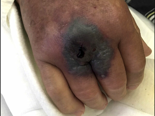
Cutaneous mucormycosis
Who gets zygomycosis?
Zygomycosis due to Mucorales is seen predominantly in immunocompromised individuals. Diabetes mellitus is the leading predisposing cause worldwide, followed by haematological malignancies and stem cell transplants. Other associations include immune compromise due to solid organ cancer or transplant, drug-induced immunosuppression, or HIV (rare), and iron overload. In up to 20% of cases, the patient is immunocompetent and infection may complicate impairment of the skin barrier such as following skin trauma, thermal burns, or toxic epidermal necrolysis, and has been reported in premature infants. Graft-transmission of infection is an important route of acquisition in some countries.
The annual incidence ranges from approximately 1/million in the US and Europe to 140/million in India.
COVID-19-associated infection with Mucorales (‘black fungus’) has particularly affected diabetic males treated with systemic corticosteroids in India during the second wave of COVID-19 (April to July 2021), resulting in over 45000 cases and at least 4000 deaths.
Entomophthorales skin infection occurs in healthy individuals particularly around the tropical and subtropical humid areas such as in equatorial Africa and Southeast Asia.
What causes zygomycosis?
Mucorales
The order Mucorales is divided into many families of which the two major causes of infection are Mucoraceae and Cunninghamellaceae. Within these families there are three important genera that cause infection in humans — Rhizopus, Mucor, and Lichtheimia (formerly Absidia) account for 75% of infections. Mucormycosis is the opportunistic infection caused by members of the Mucoraceae family.
Primary cutaneous infection generally occurs due to direct implantation of fungal elements following major or minor skin trauma, such as skin abrasion, or tattoo, or in hospital following surgery, IV-line placement, or contaminated wound dressing. Secondary cutaneous involvement follows haematogenous dissemination.
Entomophthorales
Entomophthorales are insect pathogens. Two genera of the order Entomophthorales cause subcutaneous phycomycosis in humans, Basidiobolus and Conidiobolus.
What are the clinical features of zygomycosis?
Mucormycosis
There are five major clinical forms of mucormycosis:
- Rhinocerebral (34-39%)
- Pulmonary (20-24%)
- Cutaneous (19-22%)
- Gastrointestinal
- Disseminated
- Other rare forms.
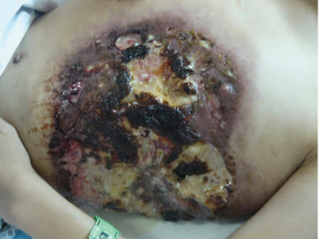
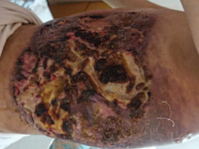
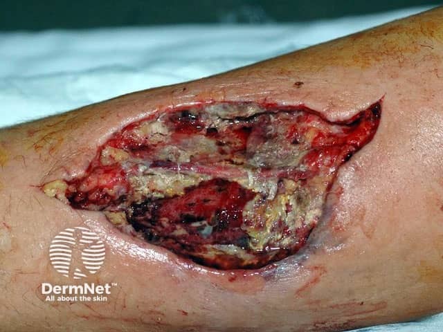
Cutaneous mucormycosis may be primary at the site of inoculation, or secondary in disseminated mucormycosis. Onset may be insidious or fulminant — acute and rapid in nature leading to gangrene.
- Necrotic eschar with erythema and induration
- Tender indurated large plaque
- Blistering and ulceration have been described
- May resemble ecthyma gangrenosum, necrotising fasciitis, pyoderma gangrenosum, or tinea corporis
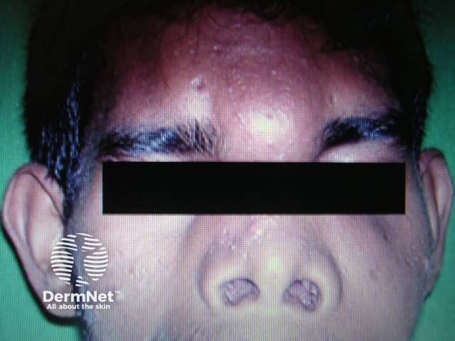
'Red flag' warning signs for rhinocerebral mucormycosis in an at-risk patient include:
- Cranial nerve palsy
- Double vision
- Sinus pain
- Protruding eye (exophthalmos)
- Swelling around an eye
- Orbital apex syndrome — vision loss, inability to move the eye
- Black necrotic ulcer on the palate.
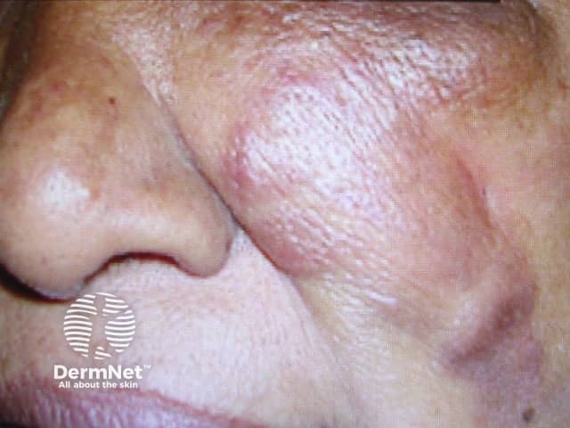
Entomophthoromycosis/Subcutaneous phycomycosis
- Slowly progressive painless subcutaneous swelling
- Uniform, disc-shaped moveable lump under the skin
- Hard, non-pitting consistency
- Typically, solitary but can be multiple
- Overlying skin usually normal
- Hyperpigmentation, peeling sometimes seen
Conidiobolus coronatus/incongruous |
|
Basidiobolus ranarum |
|
What are the complications of zygomycosis?
- Haematogenous dissemination from primary site of infection
- Invasion of adjacent tissues
- Disfigurement particularly of the face
How is cutaneous zygomycosis diagnosed?
Diagnosis of cutaneous zygomycosis may be suspected clinically in an at-risk patient and confirmed on deep incisional skin biopsy with sufficient tissue for histology and culture.
Mucormycosis
- Histology
- Eosinophilic thick-walled nonseptate hyphae
- Vasculotropic — hyphae invade vessel walls
- See Mucormycosis pathology
- Fungal culture
- Negative in up to 50%
- Allows identification of the genus and species, and antifungal agent susceptibility
Wet mount direct microscopy is a quick and inexpensive diagnostic test for mucormycosis on samples from usually sterile body tissues.
Entomophthoromycosis/Subcutaneous phycomycosis
- Histology
- Broad aseptate hyphae
- Granulomatous inflammation — lymphocytes, plasma cells, histiocytes, eosinophils, giant cells
- Splendore-Hoeppli phenomenon — smudgy eosinophilic sleeves around the hyphae
- Scattered abscesses, areas of necrosis
- Lobular panniculitis
- Fungal culture
- Fast growing fungi at 30C
- Conidiobolous species — white to greasy waxy colonies
- Basidiobolous species — cream to yellow waxy colonies
What is the differential diagnosis for zygomycosis?
What is the treatment for zygomycosis?
- Early suspicion of diagnosis
- Reversal of underlying predisposing factors
- Surgical excision — early extensive surgical debridement
- Appropriate antifungal therapy — needs to be started early within days of first symptoms, and typically continues for many months
Mucormycosis
- Amphotericin B (lipid-formulation or conventional) — high-dose IV — firstline therapy
- Posaconazole — oral or IV — used if intolerant to amphotericin B or as maintenance therapy
Entomophthormycosis
- Potassium iodide — oral
- Itraconazole — oral
What is the outcome for zygomycosis?
Zygomycosis can be difficult to treat despite the development of new antifungal agents. Treatment should be started within 6 days of symptom-onset for the most favourable outcome. Mucormycosis is often fatal although primary cutaneous disease has a lower mortality rate than gastrointestinal or pulmonary infection. In the past mortality rates were as high as 80% and even now the 90-day mortality rate is around 40%. Disseminated mucormycosis still has a mortality rate of over 90%. Rhinocerebral zygomycosis in solid organ transplant recipients is almost always fatal due to the high rate of CNS invasion.
Spontaneous resolution of entomophthormycosis has been reported. Recurrence is common even after surgical excision. The course is typically indolent.
Bibliography
- Balaji SM. Treatment algorithm for orofacial mucormycosis. Ann Maxillofac Surg. 2021;11(1):1–2. doi:10.4103/ams.ams_98_21. Journal
- Jeong W, Keighley C, Wolfe R, et al. The epidemiology and clinical manifestations of mucormycosis: a systematic review and meta-analysis of case reports. Clin Microbiol Infect. 2019;25(1):26–34. doi:10.1016/j.cmi.2018.07.011. Journal
- Jeong W, Keighley C, Wolfe R, et al. Contemporary management and clinical outcomes of mucormycosis: a systematic review and meta-analysis of case reports. Int J Antimicrob Agents. 2019;53(5):589–97. doi:10.1016/j.ijantimicag.2019.01.002. PubMed
- Kennedy KJ, Daveson K, Slavin MA, et al. Mucormycosis in Australia: contemporary epidemiology and outcomes. Clin Microbiol Infect. 2016;22(9):775–81. doi:10.1016/j.cmi.2016.01.005. Journal
- Lanternier F, Sun H-Y, Ribaud P, Singh N, Kontoyiannis DP, Lortholary O, Mucormycosis in organ and stem cell transplant recipients, Clinical Infectious Diseases. 2012; 54(11):1–8. https://doi.org/10.1093/cid/cis195. Journal
- Mendoza L, Vilela R, Voelz K, Ibrahim AS, Voigt K, Lee SC. Human fungal pathogens of mucorales and entomophthorales. Cold Spring Harb Perspect Med. 2014;5(4):a019562. doi:10.1101/cshperspect.a019562. Journal
- Pal R, Singh B, Bhadada SK, et al. COVID-19-associated mucormycosis: an updated systematic review of literature. Mycoses. 2021;10.1111/myc.13338. doi:10.1111/myc.13338. Journal
- Santosh ABR, Muddana K, Bakki SR. Response to commentary: Fungal infections of oral cavity: diagnosis, management, and association with COVID-19 [published online ahead of print, 2021 Sep 14]. SN Compr Clin Med. 2021;1–2. doi:10.1007/s42399-021-01033-9 Journal
- Skiada A, Pavleas I, Drogari-Apiranthitou M. Epidemiology and diagnosis of mucormycosis: an update. J Fungi (Basel). 2020;6(4):265. doi:10.3390/jof6040265. Journal
- Sun HY, Forrest G, Gupta KL, et al. Rhino-orbital-cerebral zygomycosis in solid organ transplant recipients. Transplantation. 2010;90(1):85–92. doi:10.1097/tp.0b013e3181dde8fc. Journal
On DermNet
- Ecthyma gangrenosum
- Fungal skin infections
- Hyperbaric oxygen
- Introduction to fungal infections
- Key clinical-trial evidence for posaconazole
- Mucormycosis pathology
- Oral antifungal medication
- Skin manifestations of systemic mycoses
- Stomatitis
- Trigeminal trophic syndrome