

Periorificial dermatitis in children — extra information
Introduction Demographics Causes Clinical features Diagnosis Differential diagnoses Treatment Outcome
What is periorificial dermatitis?
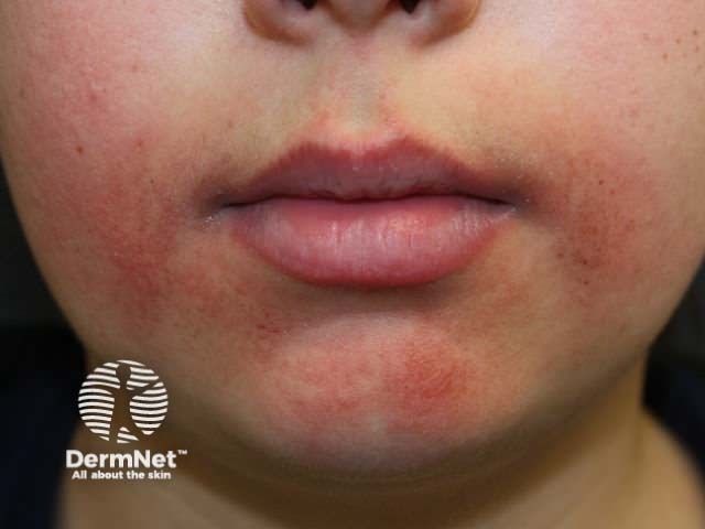
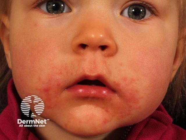
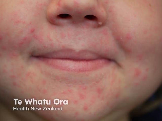
Periorificial dermatitis in children typically presents as multiple small papules around the mouth, nose, and eyes.
While the name is suggestive of an eczematous condition, periorificial dermatitis is actually more like rosacea.
Who gets periorificial dermatitis?
Periorificial dermatitis mostly affects women aged 16–45 years; it is less common in men [1,2]. It can also affect children as young as 3 months of age, with the average age of children being 6.6 years [3]. Periorificial dermatitis is slightly more common in girls than in boys and is seen most frequently in children who have had topical steroids to the face [3,4].
What causes periorificial dermatitis?
The cause of periorificial dermatitis is poorly understood.
- Children with periorificial dermatitis commonly have a background of atopic dermatitis; impaired skin barrier function may increase the impact of external irritants on the skin [5].
- Corticosteroid exposure has been noted in 58–72% of paediatric cases [3,4], including topical [3,4,6], oral [7], and inhaled corticosteroids [8]. It is thought this may be due to damage to the epidermis [2], interaction with collagen synthesis [9], or a change in follicular flora [10]. It is unclear whether corticosteroids induce periorificial dermatitis or exacerbate the pre-existing disease.
- Fluorinated dental care products, dental fillings, cosmetics, sunscreens, chewing gum, and hormonal changes have been associated with periorificial dermatitis [1,11].
- Fusobacterium and Candida albicans may also contribute [12].
What are the clinical features of periorificial dermatitis?
Periorificial dermatitis is characterised by multiple grouped erythematous papules, pustules, or vesicles, with or without any scale.
- The papules usually occur around the mouth, sparing the narrow area adjacent to the vermillion border of the lip. Other areas of the face, including around the nose and eyes, may also be involved.
- Sometimes there is itch, tenderness, or a burning sensation [11].
- Unlike acne, there are no comedones [11].
Granulomatous periorificial dermatitis is a variant mainly reported in dark-skinned prepubertal children in which there are multiple small discrete flesh-coloured or hyperpigmented papules. Granulomatous periorificial dermatitis is mainly reported in dark-skinned prepubertal children.
- The papules occur on the face and sometimes on other sites.
- In contrast to the usual type of periorificial dermatitis, erythema, papulopustules, and papulovesicles are absent [13,14].
- Granulomatous periorificial dermatitis is sometimes associated with blepharitis or conjunctivitis [15,16].
How is periorificial dermatitis diagnosed?
Periorificial dermatitis is usually diagnosed from a typical patient history and clinical features.
A skin biopsy is rarely required but may distinguish periorificial dermatitis from other disorders when the diagnosis is unclear.
- Histopathology often shows a non-specific inflammation with perifollicular lymphohistiocytic infiltrate, epithelioid cells, and occasionally giant cells [17]. Early papular lesions may demonstrate mild acanthosis, epidermal oedema, and parakeratosis [17].
- Non-caseating perifollicular granulomas are seen in granulomatous periorificial dermatitis [15].
Swabs, skin scrapings, and potassium hydroxide preparation may exclude bacterial or fungal infection.
Patch testing may be indicated if a contact allergy is suspected.
Blood tests are not useful in the diagnosis of periorificial dermatitis.
What is the differential diagnosis for periorificial dermatitis?
The differential diagnosis for non-granulomatous periorificial dermatitis in prepubertal children includes [11,18]:
- Acne in childhood
- Irritant contact dermatitis (or less often, allergic contact dermatitis)
- Atopic dermatitis
- Demodex, Candida, or fusiform infection
- Angiofibromas (a sign of tuberous sclerosis)
- Metabolic disorders, including acrodermatitis enteropathica and acrodermatitis enteropathica-like conditions
- Shwachman syndrome, and biotin deficiency.
In older children, the following conditions should also be considered:
The differential diagnosis for the granulomatous variant includes [18]:
- Sarcoidosis
- Benign cephalic histiocytosis
- Granulosis rubra nasi
- Lupus miliaris disseminatus faciei.
What is the treatment for periorificial dermatitis?
Suspected causative agents, such as topical corticosteroids, should be discontinued [1].
- Periorificial dermatitis can temporarily flare when topical corticosteroids are ceased but may subsequently resolve within a few months without additional treatment [11].
- The indication for inhaled or oral steroids should be reviewed [3]. Inhalers should be wiped clean to minimise steroid exposure.
- Skin products that may irritate or occlude the skin should also be avoided.
Mild periorificial dermatitis may be treated with a topical antibiotic, such as metronidazole, clindamycin, erythromycin and sulfacetamide [3,4,11,19]. Clearance typically takes 3 to 8 weeks [11].
Refractory or moderate to severe disease is treated with oral antibiotics.
- The typical course of treatment is 4–8 weeks [1]
- Oral tetracyclines should be avoided in children under 12 years of age [1,11].
- In younger children, erythromycin, azithromycin, and clarithromycin are used [3,4].
Other treatments reported to be effective for periorificial dermatitis include:
- 1% topical ivermectin [20]
- 20% azelaic acid cream [6]
- Topical tacrolimus [14] and pimecrolimus [21]
- Adapalene gel [22].
What is the outcome for periorificial dermatitis?
Periorificial dermatitis in children is generally benign and self-limiting and often improves spontaneously within 2–3 weeks.
- Periorificial dermatitis has been reported to resolve in 72% of children in an average time of 3.8 months [3].
- Corticosteroid use may prolong the disease course [4].
- Lesions typically resolve without scarring; however, pigmentary changes may occur [3].
- Recurrence in children is common in those who are dependent on a corticosteroid [3].
References
- Tempark T, Shwayder TA. Perioral dermatitis: A review of the condition with special attention to treatment options. Am J Clin Dermatol 2014; 15: 101–13. DOI: 10.1007/s40257-014-0067-7. PubMed
- Wollenberg A, Bieber T, Dirschka T, et al. Perioral dermatitis. J Dtsch Dermatol Ges 2011; 9: 422–7. DOI: 10.1111/j.1610-0387.2010.07329.x. PubMed
- Goel NS, Burkhart CN, Morrell DS. Pediatric periorificial dermatitis: clinical course and treatment outcomes in 222 patients. Pediatr Dermatol 2015; 32: 333–6. DOI: 10.1111/pde.12534. PubMed
- Nguyen V, Eichenfield LF. Periorificial dermatitis in children and adolescents. J Am Acad Dermatol 2006; 55: 781. DOI: 10.1016/j.jaad.2006.05.031. PubMed
- Dirschka T, Szliska C, Jackowski J, Tronnier H. Impaired skin barrier and atopic diathesis in perioral dermatitis. J Dtsch Dermatol Ges 2003; 1: 199–203. PubMed
- Jansen T, Melnik BC, Schadendorf D. Steroid-induced periorificial dermatitis in children — clinical features and response to azelaic acid. Pediatr Dermatol 2010; 27: 137–42. DOI: 10.1111/j.1525-1470.2009.00979.x. PubMed
- Clementson B, Smidt AC. Periorificial dermatitis due to systemic corticosteroids in children: report of two cases. Pediatr Dermatol 2012; 29: 331. DOI: 10.1111/j.1525-1470.2011.01651.x. PubMed
- Peralta L, Morais P. Perioral dermatitis — the role of nasal steroids. Cutan Ocul Toxicol 2012; 31: 160–3. DOI: 10.3109/15569527.2011.621918. PubMed
- Gupta R, Fonacier LS. Adverse effects of nonsystemic steroids (inhaled, intranasal and cutaneous): a review of the literature and suggested monitoring tool. Curr Allergy Asthma Rep 2016; 16: 44. DOI 10.1007/s11882-016-0620-y. PubMed.
- Takiwaki H, Tsuda H, Arase S, Takechi H. Differences between intrafollicular microorganism profiles in perioral and seborrhoeic dermatitis. Clin Exp Dermatol 2003; 28: 531–4. PubMed
- Kellen R, Silverberg B. Pediatric periorificial dermatitis. Cutis 2017; 100: 385–8. PubMed
- Hall CS, Reichenberg J. Evidence based review of perioral dermatitis therapy. G Ital Dermatol Venereol 2010; 145: 433–44. PubMed.
- Kim YJ, Shin JW, Lee JS, Park YL, Whang KU, Lee SY. Childhood granulomatous periorificial dermatitis. Ann Dermatol 2011; 23: 386–8. DOI: 10.5021/ad.2011.23.3.386. PubMed.
- Hussain W, Daly BM. Granulomatous periorificial dermatitis in an 11-year-old boy: dramatic response to tacrolimus. J Eur Acad Dermatol Venereol 2007; 21: 137–9. DOI: 10.1111/j.1468-3083.2006.01820.x. PubMed
- Urbatsch AJ, Frieden I, Williams ML, Elewski BE, Mancini AJ, Paller AS. Extrafacial and generalized granulomatous periorificial dermatitis. Arch Dermatol 2002; 138: 1354. PubMed
- Gutte R, Holmukhe S, Garg G, Kharkar V, Khopkar U. Childhood granulomatous periorificial dermatitis in children with extra-facial involvement. Indian J Dermatol Venereol Leprol 2011; 77: 703–6. DOI: 10.4103/0378-6323.86487. PubMed
- Ljubojević S, Lipozencić J, Turcić P. Perioral dermatitis. Acta Dermatovenerol Croat 2008; 16: 96–100. PubMed
- Suh KY, Frieden IJ. Perioral dermatitis. In: Irvine AD, Hoeger PH, Yan AC (eds). Harper's textbook of paediatric dermatology, 3rd edn. Hoboken, NJ: Wiley-Blackwell, 2011: 38.1–38.4.
- Green B, Morell DS. Persistent facial dermatitis: pediatric perioral dermatitis. Pediatr Ann 2007; 36: 796–8. PubMed
- Noguera-Morel L, Gerlero P, Torrelo A, Hernández-Martín Á. Ivermectin therapy for papulopustular rosacea and periorificial dermatitis in children: A series of 15 cases. J Am Acad Dermatol 2017; 76: 567–70. DOI: 10.1016/j.jaad.2016.10.034. PubMed
- Schwarz T, Kreiselmaier I, Bieber T, et al. A randomized, double-blind, vehicle-controlled study of 1% pimecrolimus cream in adult patients with perioral dermatitis. J Am Acad Dermatol 2008; 59: 34–40. DOI: 10.1016/j.jaad.2008.03.043. PubMed
- Jansen T. Perioral dermatitis successfully treated with topical adapalene. J Eur Acad Dermatol Venereol 2002; 16: 175–7. PubMed
On DermNet
- Periorificial dermatitis
- Periorificial dermatitis images
- Facial rashes
- Acne and other follicular disorders
- Steroid acne
- Rosacea
- Steroid rosacea
- Topical steroid
Other websites
- Perioral dermatitis — Australasian College of Dermatologists
- Pediatric periorificial dermatitis — Cutis journal [pdf]