DermNet provides Google Translate, a free machine translation service. Note that this may not provide an exact translation in all languages

Translate

Skin signs of respiratory disease

Author: Dr Kate Dear, Senior House Officer, St Mary’s Hospital, London, United Kingdom. DermNet New Zealand Editor in Chief: A/Prof Amanda Oakley, Dermatologist, Hamilton, New Zealand. Copy edited by Gus Mitchell, November 2017.


toc-icon
Table of contents
arrow-right-small

What is a respiratory disease?

A respiratory disease refers to any condition that affects the upper respiratory tract, trachea, lungs, and the nerves and muscles associated with breathing. The correct recognition of cutaneous signs of respiratory disease can aid clinicians in diagnosing and treating these potentially life-threatening diseases.

What are the skin signs of respiratory disease?

Cyanosis

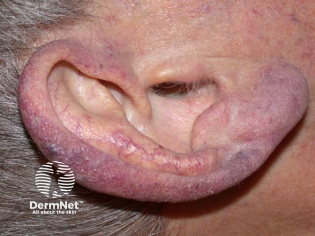
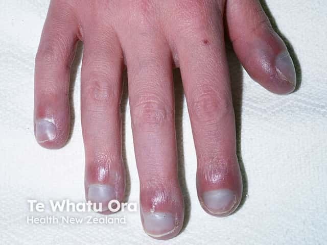
Cyanosis is a blue discolouration of the skin and mucous membranes. It is seen in patients with more than 5 g/dL of desaturated haemoglobin. Cyanosis may be:

  • Central — on the lips and tongue; relates to a circulatory or respiratory problem associated with poor blood oxygenation in the lungs
  • Peripheral — on the extremities or fingers; due to oxygen-depleted peripheral blood.

Cyanosis

Nail clubbing

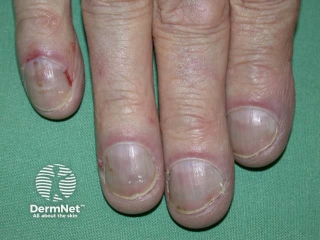
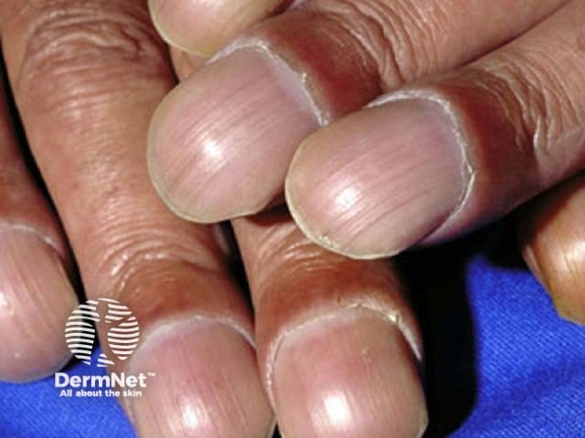
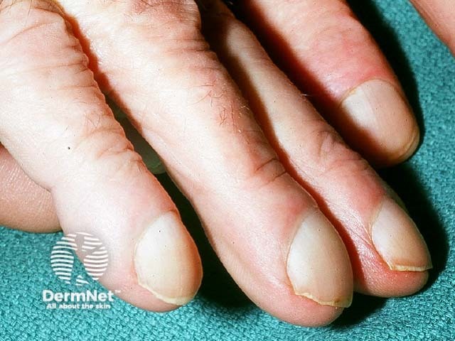
Nail clubbing is a deformity of the nails characterised by:

  • Loss of the Lovibond angle (the normal < 165° angle between the nail bed and the nail fold)
  • Thickening of the end of the finger
  • An increase in nail fold convexity
  • A soft boggy nail texture [1]. 

Clubbing is associated with a wide range of diseases, including the following respiratory diseases:

  • Lung cancer
  • Interstitial lung disease; most commonly, fibrosing alveolitis (inflammation of the alveoli in the lungs)
  • Tuberculosis
  • Cystic fibrosis
  • Suppurative (pus-forming) lung disease, such as lung abscess, empyema (pus buildup in the pleural cavity), and bronchiectasis (damage to the lungs due to infections)
  • Mesothelioma (a tumour of the lining of the lung)
  • Arteriovenous fistula or malformation (abnormal formation or connection between arteries and veins).

Hypertrophic osteoarthropathy

Hypertrophic osteoarthropathy comprises:

  • Clubbing of the fingers
  • Periostitis (inflammation of the layer of connective tissue around a bone)
  • Arthritis (inflammation of a joint).

Symptoms of hypertrophic osteoarthropathy may precede the diagnosis of lung cancer and are most commonly associated with non-small-cell lung cancer, particularly squamous cell carcinoma and adenocarcinoma [2]. Hypertrophic osteoarthropathy can also be secondary to a lung abscess, mesothelioma, and other disorders [1].

Clubbing

Lung cancer

Cutaneous metastases in lung cancer are found in 2–8% of cases of lung cancer [1]. The most common sites are the chest and abdominal wall, neck, and scalp. They are usually rapidly growing, hard, painless and mobile nodules. A punch biopsy of a nodule may be nonspecific or may establish the diagnosis of lung cancer.

Monitoring changes in the size of skin nodules can help assess the patient's response to chemotherapy [1]. Other cutaneous manifestations of lung cancer include:

Cutaneous signs of lung cancer

Superior vena cava syndrome

Superior vena cava syndrome is due to obstruction of the superior vena cava, the vein that takes deoxygenated blood from the upper body to the heart. The majority of cases are due to malignancy; most commonly, non-small-cell lung cancer, small-cell lung cancer, lymphoma, and metastatic tumours [4]. Superior vena cava syndrome presents with markedly dilated veins or venules in the upper chest [5]. These occur as a result of increased collateral flow through the superficial vasculature in the chest wall.

Sarcoidosis

Sarcoidosis can be divided into two separate conditions:

  1. A multisystem disease, with 30% of patients having cutaneous involvement [6]
  2. Cutaneous sarcoidosis, in which there is no systemic involvement [7].

Cutaneous involvement is classified as either specific, in which there are non-caseating granulomas (mixed chronic inflammatory cells) on histopathology, or nonspecific, in which granulomas are absent.

Specific lesions of sarcoidosis include:

Non–specific lesions of sarcoidosis include:

Cutaneous sarcoidosis

Cystic fibrosis

Cutaneous manifestations of cystic fibrosis may be nonspecific, but include:

  • Aquagenic skin wrinkling, due to increased concentration of electrolytes in the sweat
  • Cutaneous vasculitis, due to circulating antigen-antibody complexes (the antigens are bacteria, antibiotics and pancreatic enzyme supplements)
  • Cystic fibrosis nutrient-deficiency dermatitis [9].

Cutaneous signs of cystic fibrosis

Granulomatosis with polyangiitis

Cutaneous manifestations of granulomatosis with polyangiitis (also known as Wegener granulomatosis) are found in 10% of patients at diagnosis and develop in 50% of patients throughout the disease [1,8].

Skin findings include:

Cutaneous signs of granulomatosis with polyangiitis

Eosinophilic granulomatosis with polyangiitis

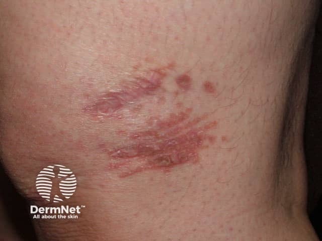
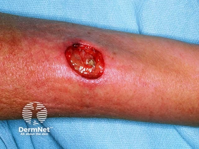
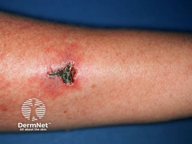
Eosinophilic granulomatosis with polyangiitis (also known as Churg-Strauss syndrome) is a rare multisystem disorder that primarily affects the lungs, skin, and peripheral nervous system. The main features are asthma, eosinophil infiltration in the lungs and blood, and small-vessel vasculitis with granulomas on biopsy.

A variety of skin lesions are seen in eosinophilic granulomatosis with polyangiitis:

Papules and nodules may become necrotic, starting with a central black dimple.

Vasculitis in eosinophilic granulomatosis

Pulmonary arteriovenous malformations

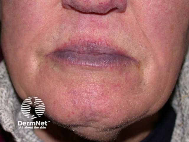
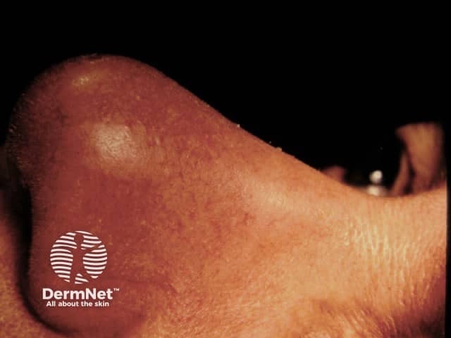
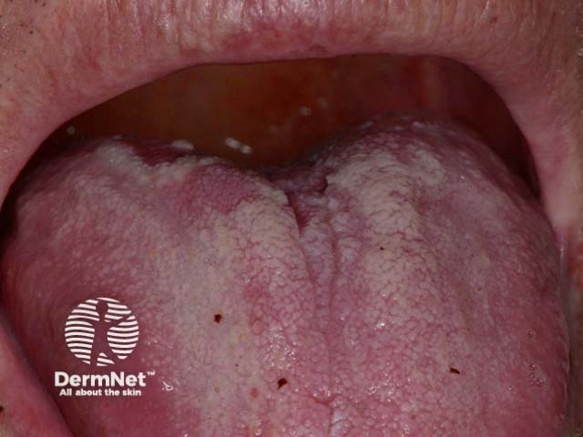
Pulmonary arteriovenous malformations are abnormal communications between the arteries and veins that supply the lungs. The majority of these malformations are congenital. Approximately 70% of patients with pulmonary arteriovenous malformations have associated hereditary haemorrhagic telangiectasia (also known as Osler–Weber–Rendu syndrome) in which telangiectasia is near the surface of the skin. The face, hands, feet, chest, lips, tongue, oral mucosa, and nasal mucosa are most commonly affected [1].

Alpha-1-antitrypsin deficiency

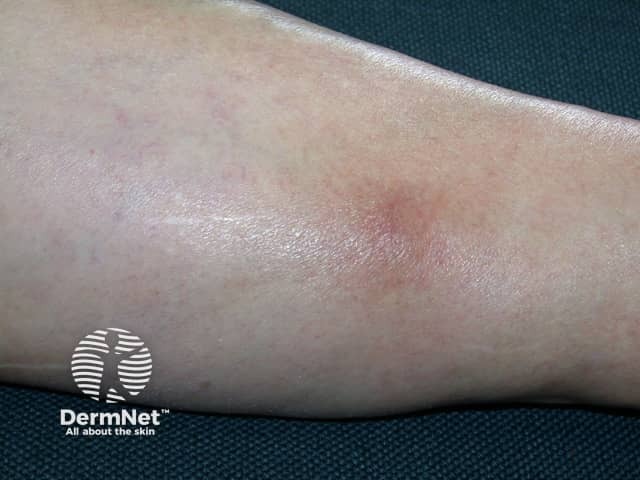
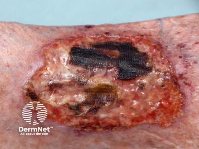
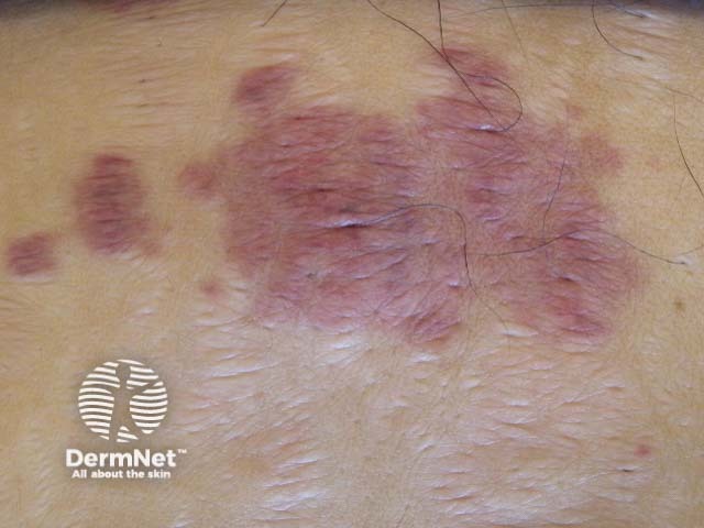
Alpha-1-antitrypsin deficiency is a genetic disorder where there is defective production of the alpha-1-antitrypsin enzyme. It primarily affects the lungs (with emphysema), liver, and skin. Cutaneous findings include recurrent ulcerative, necrotising panniculitis (inflamed subcutaneous fat with crops of painful, red subcutaneous nodules), which most noticeably occurs on the trunk and proximal extremities. These nodules typically ulcerate, releasing a clear to yellow oily fluid [1].

Fat embolism syndrome

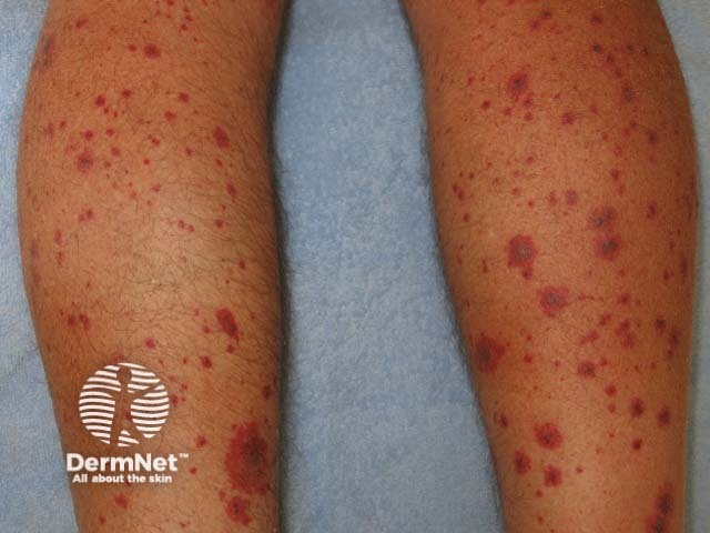
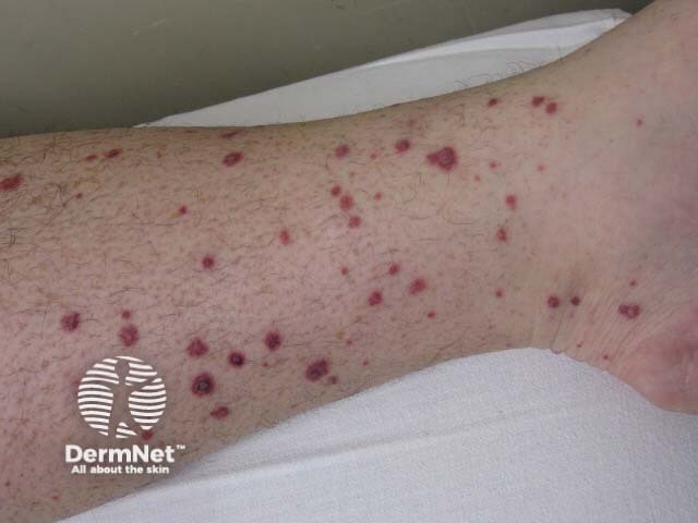
Fat embolism syndrome typically follows fractures and trauma to the long bones or pelvis. Fat particles released into the circulation may cause:

  • Respiratory distress
  • Altered mental status
  • Thrombocytopenia.

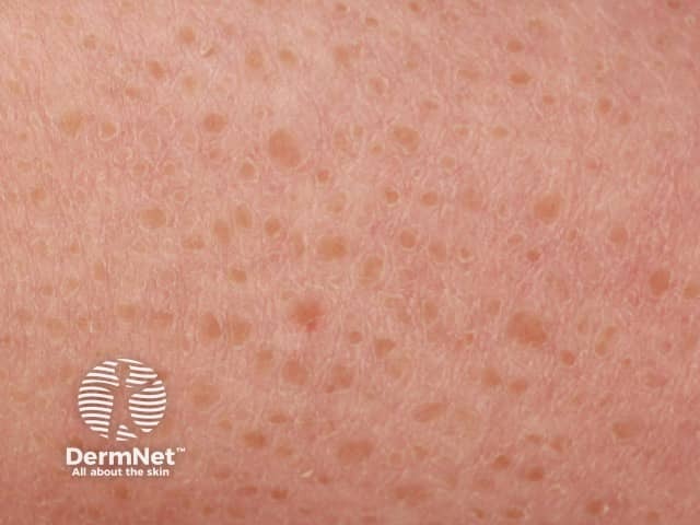
The classic cutaneous manifestation of fat embolism syndrome is a petechial eruption with 2–3 mm purpuric macules on non-dependent portions of the body (upper chest, neck, axillae, and conjunctivae). This finding often disappears within 5–7 days, but may be key for diagnosis [10,11].

Yellow nail syndrome

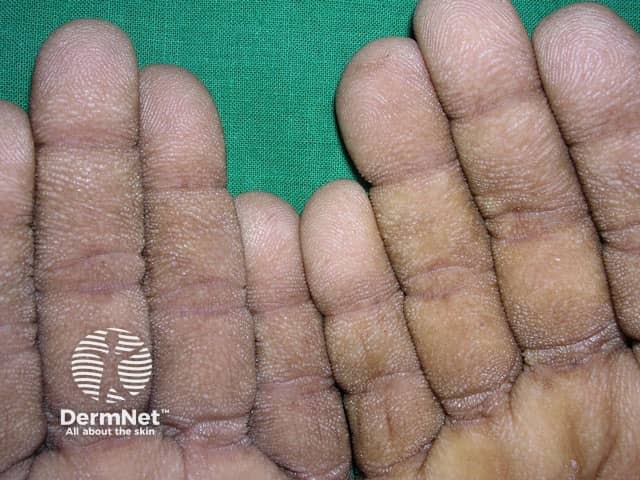
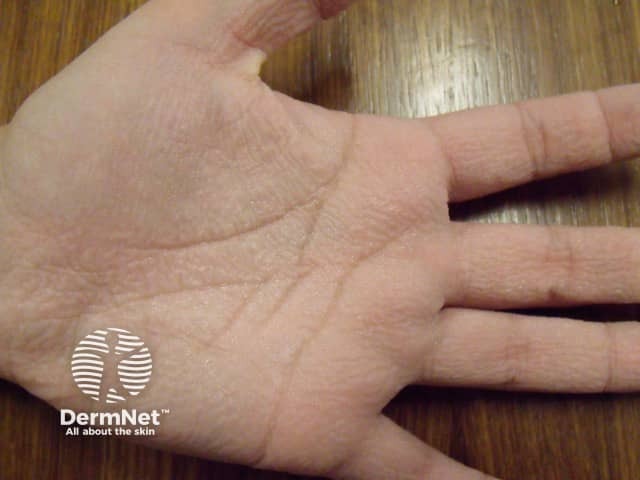
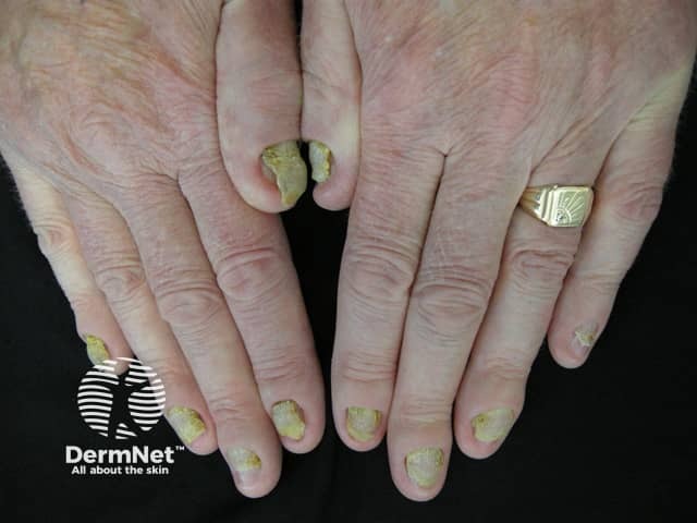
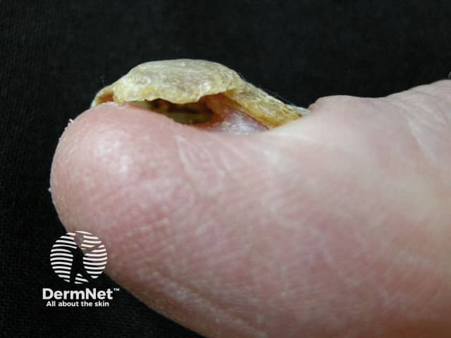
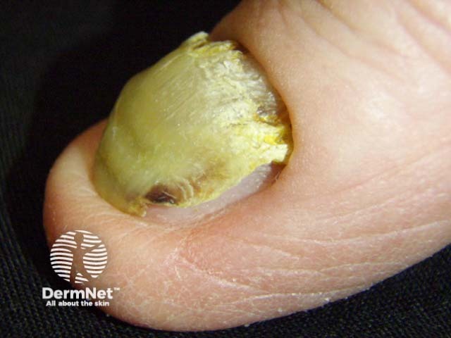
Yellow nail syndrome is the triad of:

  1. Yellow nails
  2. Lymphoedema (swelling caused by blockage of the lymphatic system)
  3. Pleural effusions (fluid in the space surrounding the lungs) [8].

Nail changes that are usually the first features of yellow nail syndrome include:

  • Onycholysis (separation of the nail from the nail bed)
  • Transverse ridging on a smooth base
  • Loss of the cuticle
  • Slow growth (less than half the rate of healthy nails) [7].

The nails are occasionally green in colour. Lymphoedema associated with yellow nail syndrome typically affects the legs and is symmetrical and non-pitting.  

Yellow nail syndrome

Birt–Hogg–Dubé syndrome

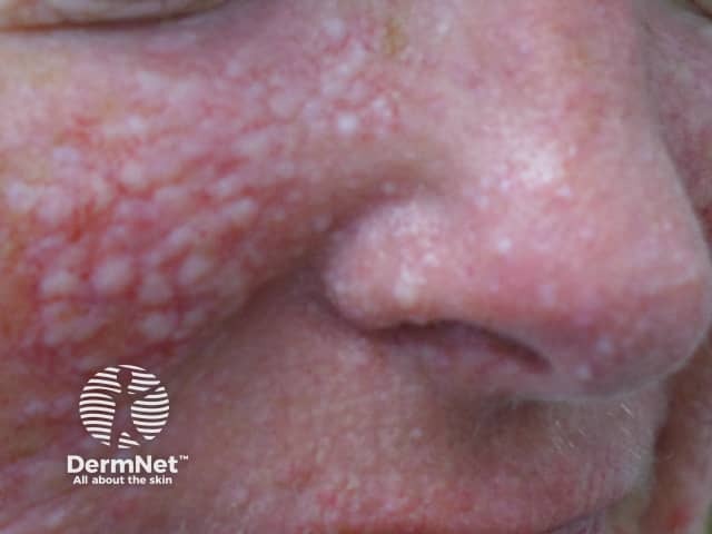
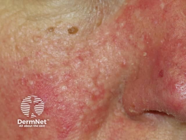
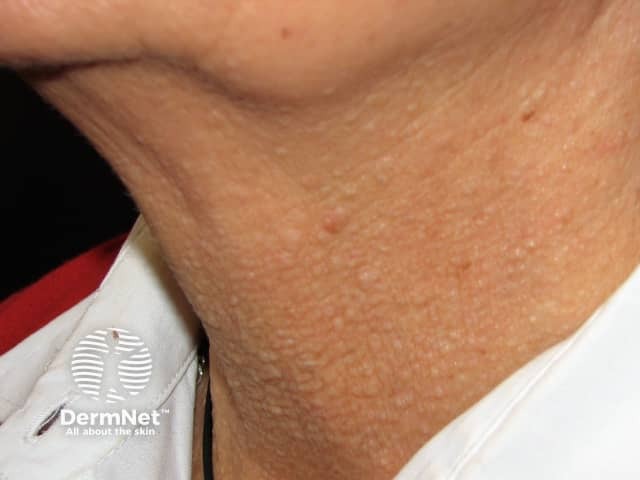
Birt–Hogg–Dubé syndrome is an autosomal-dominant inherited disease characterised by skin tumours, pneumothorax (lung collapse), kidney tumours, and lung cysts [12].

Cutaneous lesions include:

Fibrofolliculomas and trichodiscomas are most frequently found on the head, face, and upper body.

  • They are asymptomatic, pale yellow, slightly raised, dome-shaped papules measuring 2–4 mm in diameter.
  • There may be few or many hundreds of lesions.  
  • They usually develop at around 30–40 years of age [1].

Trichofolliculomas in Birt-Hogg-Dubé syndrome


References

  1. Kaldas M. Dermatologic manifestations of pulmonary disease. Medscape. January 2015. Available at: https://emedicine.medscape.com/article/1094030-overview (accessed November 2017).
  2. Davis M, Sherry V. Hypertrophic osteoarthropathy as a clinical manifestation of lung cancer. Clin J Oncol Nurs 2011; 15 (5): 561–3. DOI: 10.1188/11.CJON.561-563. PubMed
  3. Owen C. Cutaneous manifestations of lung cancer. Semin Oncol 2016; 43: 366–9. DOI: 10.1053/j.seminoncol.2016.02.025. PubMed
  4. Wilson L, Detterbeck F, Yahalom J. Clinical practice: superior vena cava syndrome with malignant causes. N Engl J Med 2007; 356: 1862–69. DOI: 10.1056/NEJMcp067190. PubMed
  5. Ratnarathorn M, Craig E. Cutaneous findings leading to a diagnosis of superior vena cava syndrome: A case report and review of the literature. Dermatol Online J 2011; 17 (6): 4. PubMed
  6. Noe M, Rosenbach M. Cutaneous sarcoidosis. Curr Opin Pulm Med 2017; 23: 482–6. DOI: 10.1097/MCP.0000000000000402. PubMed
  7. Soutor C, Hordinsky M. Clinical dermatology, 1st edn. London: McGraw Hill, 2013.
  8. Weller R. Clinical dermatology, 5th edn. Oxford: John Wiley & Sons, 2015.
  9. Rullán, J, Seijo-Montes R, Vaillant A, Sánchez N. Cutaneous manifestations of pulmonary disease. In: Sánchez N (ed). Atlas of dermatology in internal medicine. New York: Springer, 2012: 17–30.
  10. Aparicio G, Soler I, López-Durán L. Fat embolism syndrome after nailing an isolated open tibial fracture in a stable patient: a case report. BMC Res Notes 2014; 7: 237. Journal
  11. Kosova E, Bergmark B, Piazza G. Fat embolism syndrome. Circulation 2015; 131: 317–20. DOI: 10.1161/CIRCULATIONAHA.114.010835. Journal
  12. Habif T. Clinical dermatology: a colour guide to diagnosis and therapy, 6th edn. St Louis: Saunders, 2016.

On DermNet

Books about skin diseases


Related information

Sign up to the newsletter