

Autoimmune diseases in dermatology — extra information
Introduction - immune system
Introduction - autoimmunity
Demographics
Causes
Types of autoimmune blistering diseases
Diagnosis
Differential diagnoses
Treatment
What is the immune system?
The immune system is made up of the cells, molecules, and structures that defend the body from skin infections and monitor for tissue damage [1].
Immune responses can be divided into innate immunity and adaptive immunity.
Innate immunity
Innate immunity describes generalised measures to ward off infection. These include:
- Physical barriers, such as the skin and mucous membranes, which stop the majority of microorganisms entering [2]
- White blood cells, such as macrophages, which recognise microorganisms and release chemicals to stimulate and attract other immune cells
- Neutrophils, which envelope and digest invading microorganisms by a process called phagocytosis (cellular ‘eating’).
Innate immune responses are often accompanied by inflammation [3].
Adaptive immunity
Adaptive immunity describes slower immune responses, including the production of immune cells that produce specific antibodies to target and remove a particular microorganism [1].
- In cell-mediated immunity, T lymphocytes are produced that are conditioned to eliminate intracellular pathogens (viruses and bacteria) [4].
- In humoral immunity, antibody-producing B lymphocytes deal with extracellular pathogens (bacteria in a polysaccharide capsule) [4].
Adaptive immunity results in the production of memory T lymphocytes (cells that have previously encountered an antigen and have "experience" fighting infections) and B lymphocytes (which produce antibodies) that are able to specifically target a particular infection. These lymphocytes continue to circulate and quickly recognise and remove the particular virus or bacteria when they are next encountered [3].
What is autoimmunity?
Autoimmunity is an immune response against the self that usually involves T and B lymphocytes. The particular protein or structure targeted by the T and B lymphocytes is called the self-antigen [3].
Autoimmunity may result in autoimmune disease with tissue damage or impaired physiological function. Autoimmune responses may also occur without causing disease [3].
Antibodies that react against self-antigens are called autoantibodies. In some autoimmune diseases, autoantibodies are the direct cause of tissue damage. In others, autoantibodies may be present without causing injury [5].
Examples of autoimmune diseases include:
- Graves disease (a cause of hyperthyroidism)
- Rheumatoid arthritis
- Insulin-dependent diabetes
- Multiple sclerosis
- Systemic lupus erythematosus [1].
Autoimmune skin diseases
Autoimmune blistering skin conditions diseases include:
- Pemphigus vulgaris and other forms of pemphigus
- Bullous pemphigoid and other forms of pemphigoid
- Epidermolysis bullosa acquisita
- Dermatitis herpetiformis.
Autoimmune bullous diseases
Autoimmunity and deregulation of the immune system also contribute to many skin diseases, such as:
Autoimmune skin diseases
Who gets autoimmune diseases?
Autoimmune diseases affect around 5% of the population [3].
- Most autoimmune diseases are more common in women [3].
- People with a family history of autoimmune disease are at higher risk of developing an autoimmune disease themselves [6].
- Some studies show autoimmune diseases to be more common among patients from higher socioeconomic groups and northern latitudes [3].
- Autoimmune blistering skin diseases are rare and have fairly similar rates in both men and women [7].
What causes autoimmune diseases?
Autoimmune disease occurs when the responses that normally prevent autoimmunity fail [5]. There are several protective mechanisms.
- Maturing T lymphocytes in the thymus (a lymphoid organ found between the lungs where the T cells develop and mature) are removed if they react strongly against self-antigens [3].
- Circulating T-regulatory cells suppress immune responses [5].
- B lymphocytes that react strongly against self-antigens can undergo receptor editing and change their B-cell receptors [5].
- T and B lymphocytes in the circulation need co-stimulation by other immune cells to become active [5].
The exact cause of a particular autoimmune disease is often not fully understood. Risk factors for many autoimmune diseases include genetic factors, infections, hormones, and drugs.
- Genetic factors are most commonly polygenic (ie, multiple genes combine to increase risk) [1].
- Infections may trigger an autoimmune process by mimicking a self-antigen or by increasing co-stimulatory molecules [1].
- Genes on the Y chromosome may protect men from autoimmune disease, and oestrogen may play a role in the increased susceptibility of women to autoimmune diseases [1].
- Certain drugs (eg, penicillamine, captopril, and vancomycin) can precipitate pemphigus vulgaris and pemphigus foliaceus [8].
Well-designed trials have concluded that there is no evidence that vaccines cause autoimmune disease [5].
Autoimmune blistering diseases
The autoimmune cause of this class of blistering skin conditions is confirmed by positive direct immunofluorescence microscopy revealing the deposition of antibodies in the skin.
Pemphigus
Pemphigus is a group of rare blistering disorders caused by circulating autoantibodies that bind to adhesion molecules in the skin, which disrupts keratinocytes from sticking together, causing intraepidermal blisters [9]. The main types of pemphigus are pemphigus vulgaris, pemphigus foliaceus, and paraneoplastic pemphigus [9].
- Pemphigus vulgaris is characterised by blisters and erosions inside the mouth as well as on the skin. There are circulating autoantibodies against desmoglein 3, which is a protein important in keratinocyte cell-to-cell adhesion [10].
- Pemphigus foliaceus causes superficial blisters typically on the trunk, scalp, and face. There are autoantibodies against desmoglein–1, a molecule that adheres skin cells to each other [3].
- Paraneoplastic pemphigus causes blistering and ulceration in the mouth and sometimes the skin; it arises in association with malignancy, most often a non-Hodgkin lymphoma. The multiple autoantibodies seen in paraneoplastic pemphigus include those targeting desmoplakin proteins [11].
Pemphigoid
The pemphigoid group of diseases includes bullous pemphigoid, mucous membrane pemphigoid, and pemphigoid gestationis. Pemphigoid blisters are subepidermal and are caused by autoantibodies that bind in the area of the dermal-epidermal junction [9].
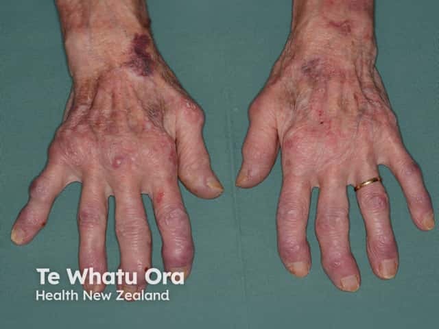
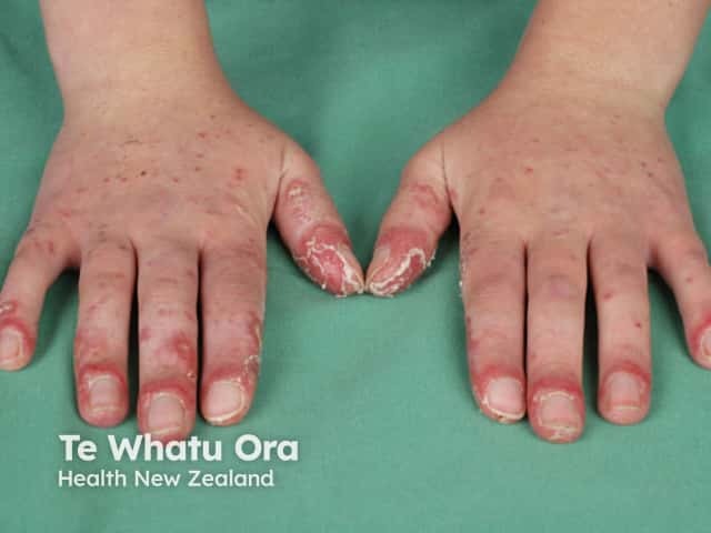
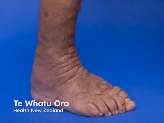
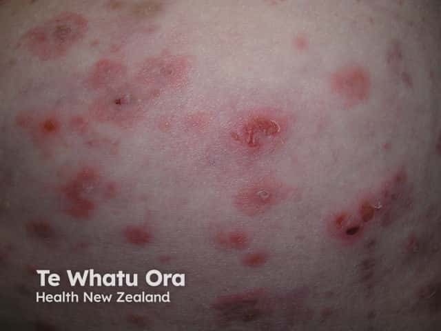
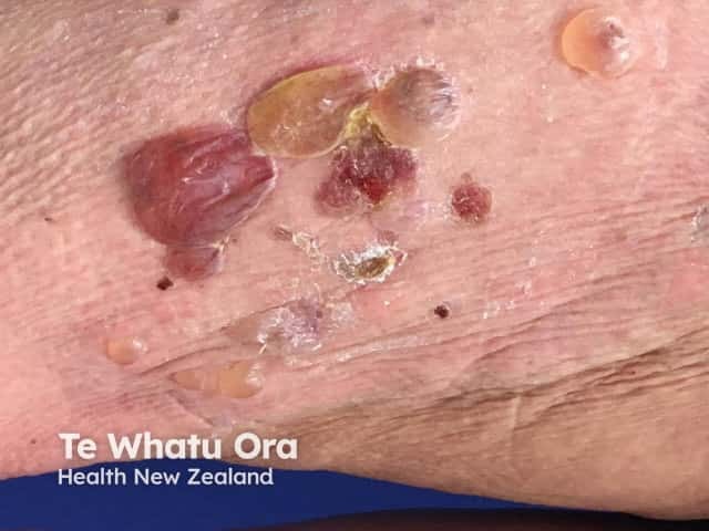
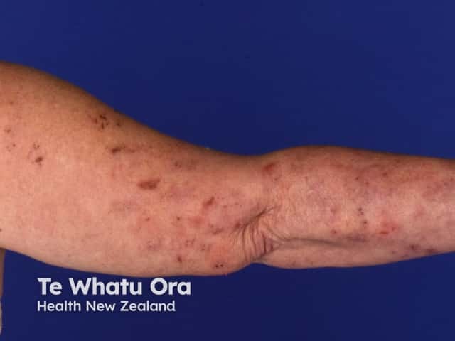
- Bullous pemphigoid predominantly affects older people who present with large tense fluid-filled blisters and erosions, often preceded by urticated or eczematous plaques [12]. There are antibodies against bullous pemphigoid antigen (BP180), a hemidesmosome-associated protein involved in keratinocyte to basement membrane adhesion, and against bullous pemphigoid antigen 230 (BP230), a protein found in basal keratinocytes [9].
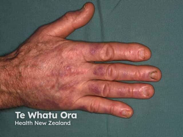
- Mucous membrane pemphigoid is characterised by recurrent blistering and ulceration of mucous membranes, particularly in the mouth and eyes, and can also affect the skin [9]. The split occurs lower in the dermal-epidermal junction, leading to scarring. Several autoantibodies have been associated with different presentations of mucous membrane pemphigoid including BP180, BP230, laminin 332, integrin alpha 6 and beta 4, and type VII collagen.
- Pemphigoid gestationis affects women during pregnancy or shortly after delivery. Pemphigoid gestationis often starts as an intensely itchy urticaria-like rash around the belly button and then may spread to involve the entire skin surface, but not mucous membranes. It later typically progresses to tense blisters resembling those of bullous pemphigoid [13]. BP180 antibodies may be detected. The primary site of autoimmunity is thought to be the placenta.
Other rare blistering diseases
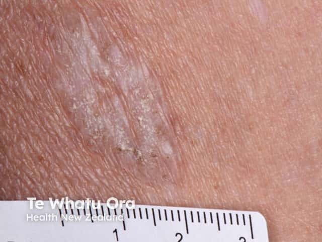
Dermatitis herpetiformis is an itchy blistering skin disease that typically affects the elbows, knees, and buttocks. It is associated with coeliac disease and the symptoms heal with a gluten-free diet [14]. It is characterised by blisters with a subepidermal deposition of immunoglobulin A (IgA) and a neutrophilic infiltrate. In dermatitis herpetiformis, the antibodies in the autoimmune response target the coagulation enzyme epidermal transglutaminase [15].
Linear IgA bullous dermatosis is a very rare autoimmune blistering disorder that can be acquired or drug-induced (eg, by vancomycin). The blisters are sometimes arranged in rings (known as the ‘pearl necklace’ sign) [9]. There is a subepidermal deposition of IgA antibodies which target a portion of the BP180 antigen, type VII collagen, or other basement membrane proteins.
Epidermolysis bullosa acquisita is also very rare. In its classical form, blisters and erosions form at areas of minor trauma [9]. In epidermolysis bullosa acquisita, the autoimmune reaction is directed against type VII collagen in the basement membrane zone of the skin and mucosa.
Bullous systemic lupus erythematosus is a rare presentation of subepidermal blistering in a patient with systemic lupus erythematosus.
How are autoimmune diseases in dermatology diagnosed?
A biopsy is usually needed for a definitive diagnosis of autoimmune skin disease, although a characteristic appearance may be suggestive of a particular condition [9].
Blood tests may include:
- Tests for circulating skin autoantibodies (indirect immunofluorescence) [14]
- Tests for coeliac antibodies, such as IgA tissue transglutaminase antibodies
- Nonspecific tests for inflammatory markers (eg, C-reactive protein)
- An autoimmune screen (eg, anti-nuclear antibodies).
Swabs may be taken of a ruptured blister to look for signs of a bacterial infection or herpes virus infection.
What is the differential diagnosis for autoimmune blistering skin diseases?
Blistering skin rashes associated with systemic illness may present with fever and ‘flu-like' symptoms [14]. These disorders can include:
- Steven–Johnson syndrome / toxic epidermal necrolysis
- Staphylococcal scalded skin syndrome
- Varicella-zoster infection
- Disseminated herpes zoster, eczema herpeticum, or herpes simplex in immunocompromised patients
- The bullous form of systemic lupus erythematosus
- Acute febrile neutrophilic dermatosis.
Other generalised blistering disorders that present with autoimmune skin disease symptoms include:
The blistering of mucous membranes is also a common symptom in these conditions:
- Erythema multiforme
- Viral skin infection (eg, herpes simplex, herpes zoster, or enteroviral stomatitis)
- Aphthous ulcers
- Behçet syndrome [14].
What is the treatment for autoimmune skin diseases?
The treatment of autoimmune skin diseases depends on the specific condition.
Pemphigus and pemphigoid are mainly treated with systemic corticosteroids and immunosuppressive treatments [16].
Dermatitis herpetiformis is treated with dapsone and a gluten-free diet [15].
References
- Delves PJ, Martin SJ, Burton DN, Roitt IM. Roitt’s essential immunology, 12th edn. Chichester: Wiley-Blackwell, 2011.
- Johnstone RB. An overview of the innate immune system. UpToDate. Updated 7 November 2017. Available at: www.uptodate.com/contents/an-overview-of-the-innate-immune-system (accessed 4 February 2018).
- Chapel H, Haeney M, Misbah S, Snowden N. Essentials of clinical immunology, 6th edn. Oxford Wiley-Blackwell, 2014.
- Maddoff LC, Kasper DL. Introduction to infectious diseases: host-pathogen interactions. In: Longo D, Fauci A, Kasper D, Hauser S, Jameson J, Losclazo J (eds). Harrison’s principles of internal medicine, 18th edn. McGraw-Hill, 2012: 1007–12.
- Rose NR. Overview of autoimmunity. UpToDate. Updated 10 July 2017. Available at: www.uptodate.com/contents/overview-of-autoimmunity (accessed 7 February 2018).
- Diamond B, Lipsky PE. Autoimmunity and autoimmune diseases. In: Longo D, Fauci A, Kasper D, Hauser S, Jameson J, Loscalzo J (eds). Harrison’s principles of internal medicine, 18th edn. New York: McGraw-Hill, 2012: 2719–24.
- Bickle KM, Roark TR, Hsu S. Autoimmune bullous dermatoses: A Review. Am Fam Physician 2002; 65: 1861–71. PubMed
- Hertle M, Sitaru C. Pathogenesis, clinical manifestations, and diagnosis of pemphigus. UpToDate. Updated 23 May 2017. Available at: www.uptodate.com/contents/pathogenesis-clinical-manifestations-and-diagnosis-of-pemphigus (accessed 4 February 2018).
- Baum S, Sakka N, Artsi O, Trau H, Brazilai A. Diagnosis and classification of autoimmune blistering diseases. Autoimmun Rev 2014; 13: 482–9. DOI: 10.1016/j.autrev.2014.01.047. PubMed
- Habif T. Vesicular and bullous diseases. In: Habif T. Clinical dermatology: a color guide to diagnosis and therapy, 5th edn. Edinburgh/New York: Mosby Elsevier, 2009: 635–70.
- Anhalt GJ. Paraneoplastic pemphigus. UpToDate. Updated 5 October 2017. Available at: www.uptodate.com/contents/paraneoplastic-pemphigus (accessed 1 March 2018).
- Yancey KB, Lawley TJ. Immunologically mediated skin diseases. In: Longo D, Fauci A, Kasper D, Hauser S, Jameson J, Loscalzo J (eds). Harrison’s principles of internal medicine, 18th edn. New York: McGraw-Hill, 2012: 424–31.
- Ambros-Rudolph CM. Dermatoses of pregnancy – clues to diagnosis, fetal risk and therapy. Ann Dermatol 2011; 23: 265–75. DOI: 10.5021/ad.2011.23.3.265. PubMed Central
- Hull C, Zone J. Approach to patient with cutaneous blisters. UpToDate. Updated 10 July 2017. Available at: www.uptodate.com/contents/approach-to-the-patient-with-cutaneous-blisters (accessed 21 April 2018).
- Collin P, Salmi TT, Hervonen K, Kaukinen K, Reunala T. Dermatitis herpetiformis: a cutaneous manifestation of celiac disease. Ann Med 2017; 49: 23–31. DOI: 10.1080/07853890.2016.1222450. PubMed
- Kayani M, Aslam AM. Bullous pemphigoid and pemphigus vulgaris. BMJ 2017; 357: j2169. DOI: 10.1136/bmj.j2169. Journal
On DermNet
Other websites
- Skin in systemic disease — DermNet e-lecture [Youtube]