Blistering skin conditions — extra information
Introduction
Acute blistering diseases
Chronic blistering diseases
E-lecture
What is a blistering disease?
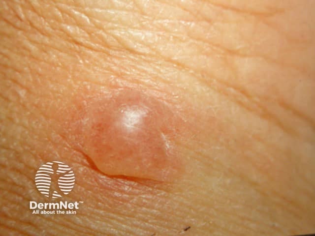
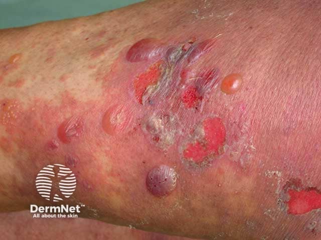
A blistering disease is a condition in which there are fluid-filled skin lesions.
- Vesicles are small blisters less than 5 mm in diameter.
- A bulla is a larger blister. Note that the plural of bulla is bullae.
- Blisters may break or the roof of the blister may become detached forming an erosion. Exudation of serous fluid forms crust.
Acute blistering diseases
Acute blistering diseases can be generalised or localised to one body site and are due to infection or inflammatory disorders. Although most commonly eczematous, generalised acute blistering diseases can be life-threatening and often necessitate hospitalisation.
Acute blistering conditions should be investigated by taking swabs for bacterial and viral culture. A skin biopsy may be helpful in making a diagnosis.
Acute generalised blistering diseases
Acute febrile neutrophilic dermatosis
- Neck, limbs, upper trunk
- Pseudovesicular plaques, blisters, pustules, purpura, or ulceration
- Disease associations: rheumatoid arthritis, inflammatory bowel disease, autoimmune arthritis, myeloid dysplasia
- Biopsy suggestive
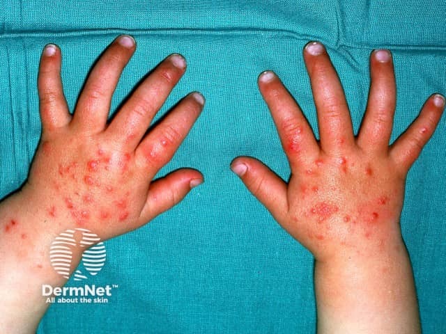
Atypical enterovirus infection
- Widespread vesicular eruption
- Clears in a few days
- When affecting areas of atopic dermatitis, enteroviral infection is called eczema coxsackium.
- Childhood illness; more serious in adults
- Scalp, face, oral mucosa, trunk
- Culture/PCR Herpes varicella zoster virus
- Traditionally linked to barbiturate use; other drugs have been implicated.
- Develop 48 - 72 hours after coma onset.
- Self-limiting, usually resolving within 1 to 2 weeks.
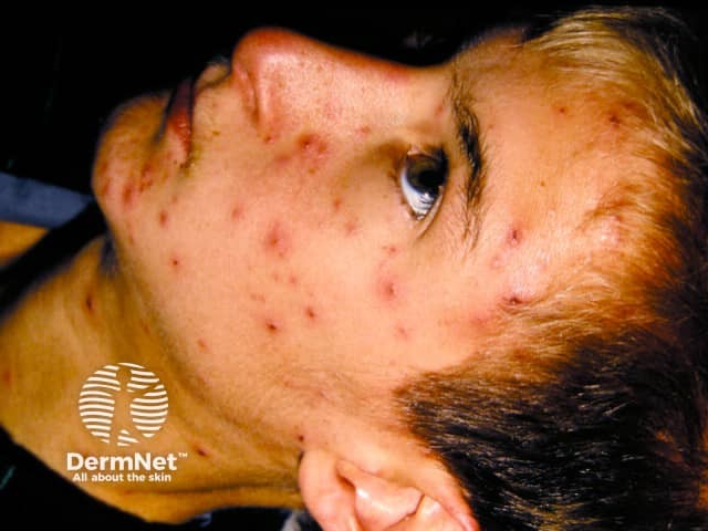
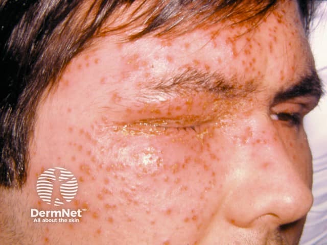
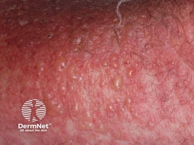
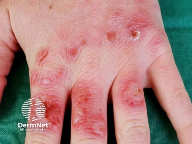
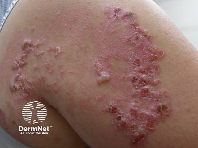
- History of atopic dermatitis
- Monomorphic cluster of umbilicated vesicles
- Culture/PCR Herpes simplex virus
- Affects body sites exposed to the sun (hands, upper chest, feet
- Papules, plaques, sometimes targetoid
- May spare face
- Arises within hours of exposure to bright sunlight
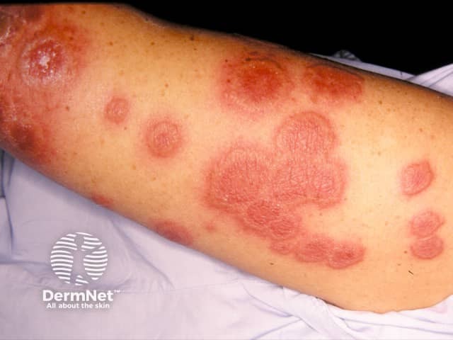
- A reaction eg, to infection
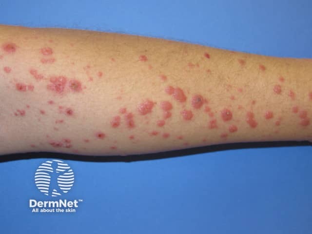
- An acute eruption of papules, plaques, target lesions
- Acral distribution: cheeks, elbows, knees, hands, feet
- May have mucositis (lips, conjunctiva, genitalia)
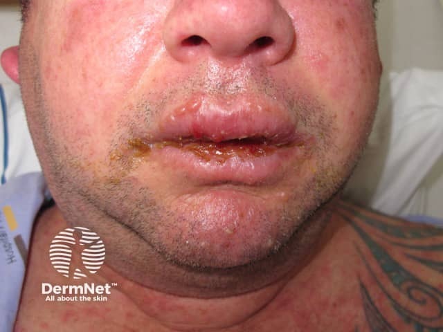
Stevens-Johnson syndrome / toxic epidermal necrolysis
- Patient very unwell
- Mucosal involvement
- Nearly always drug-induced
- Rarely due to mycoplasma infection
- Painful red skin may come off in sheets or have multiple coalescing blisters
Drug hypersensitivity syndrome
- Drug started up to 8 weeks prior to the onset
- Morbilliform eruption that may blister (without necrolysis)
- Often mucosal involvement
- Multiorgan damage (renal, hepatic, respiratory, haematological)
- Often, marked eosinophilia
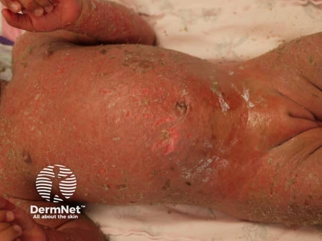
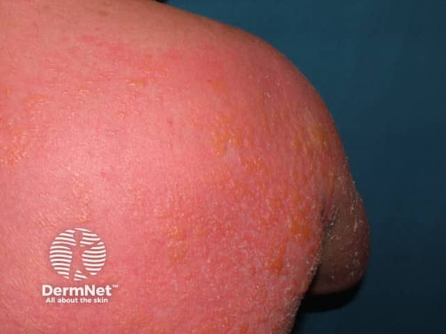
Staphylococcal scalded skin syndrome
- Young child
- Miserable
- Red skin comes off in sheets
- Evidence of staphylococcal infection
Acute localised blistering diseases
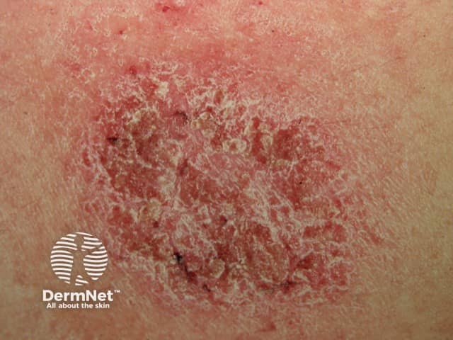
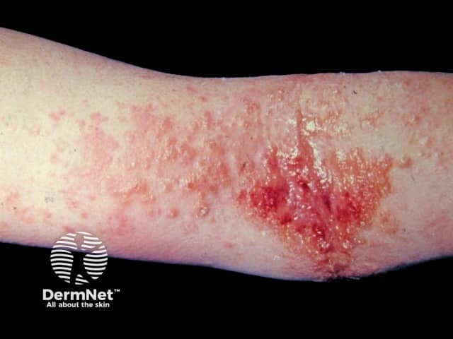
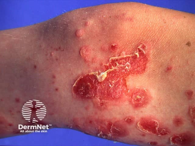
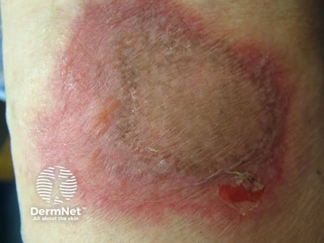
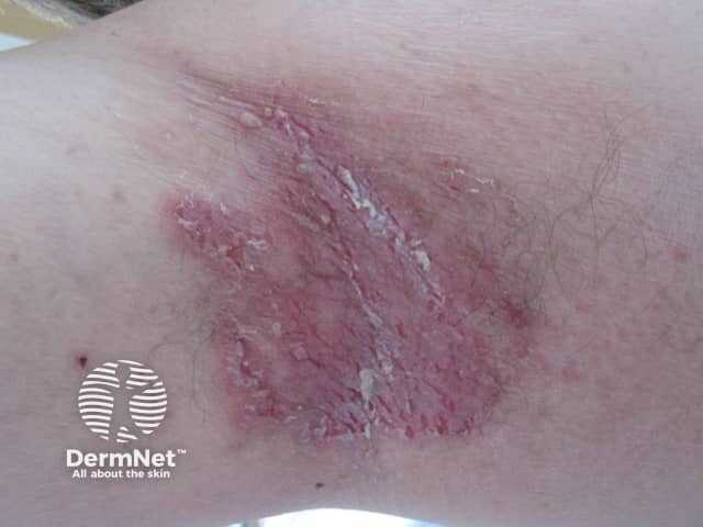
Acute dermatitis
- Rapidly enlarging plaque
- Swab Staphylococcus aureus
- Wound infections, scabies, etc
- Fingers, toes, acral sites
- Exposed to cold
- Purplish itchy/burning plaques
Enteroviral vesicular stomatitis
Clears in a few days
- Acute febrile illness
- Swab Streptococcus pyogenes
- Recurring rash, typically at the same site
- Due to an intermittent drug that has been taken within 24 hours of the rash appearing
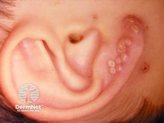
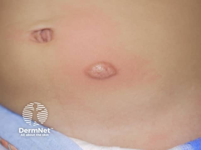
- Single or few lesions
- Central blister
- Monomorphic, umbilicated
- Culture/PCR Herpes simplex virus
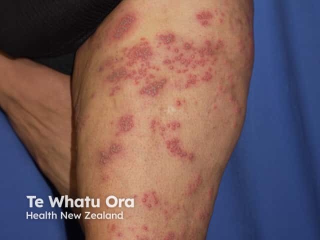
- Dermatomal distribution
- Culture/PCR Herpes varicella zoster virus
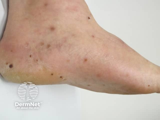
- Crops of urticated papules
- Central vesicle or punctum
- Favour exposed sites
- Blisters due to contact with various beetles (eg, paedarus dermatitism, lax beetle dermatosis) and caterpillars
- Central trunk
- Sweat rash
- Vesicles are very superficial
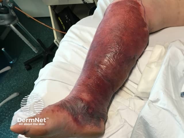
- Very sick; septic shock
- Rapid spread of cellulitis with purpura/blistering
- Anaesthetic areas in early lesions
- Bacterial culture essential
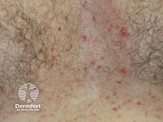
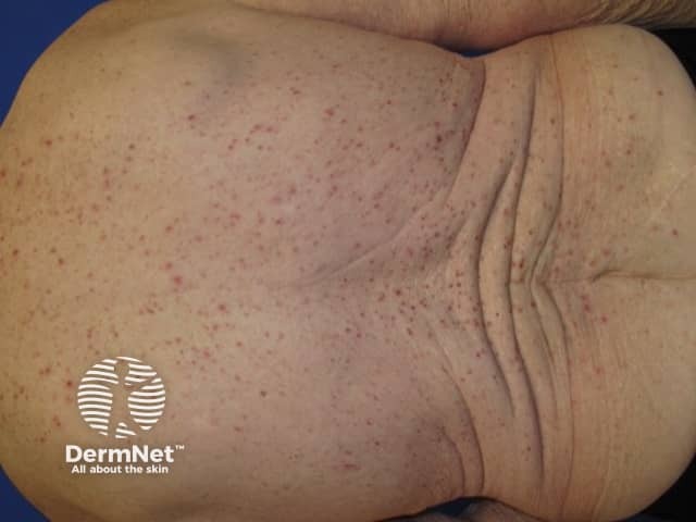
Transient acantholytic dermatosis
- Acute or chronic
- Elderly males
- Itchy or asymptomatic
- Crusted papules
- Extremely rare autosomal recessive genodermatosis
- Form of generalised peeling skin syndrome
- May be accompanied by blistering
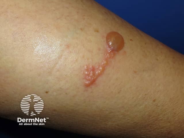
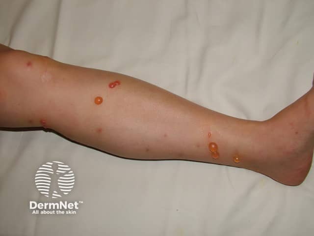
Trauma
- History of injury or neuropathy
- Friction, thermal burns, ultraviolet radiation sunburns, chemical burns, fracture
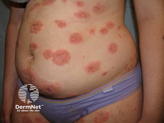
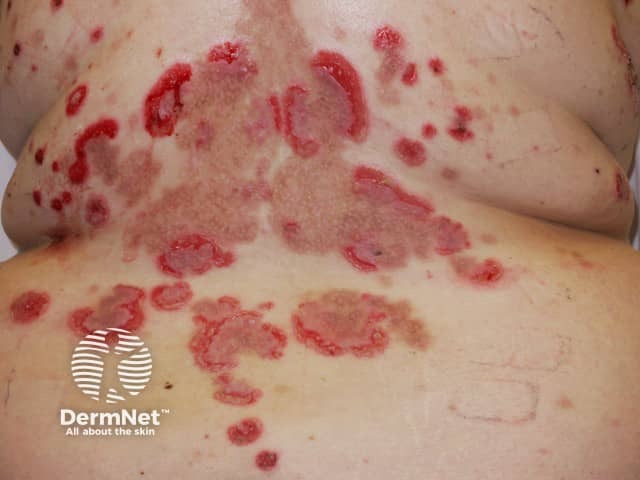
Chronic blistering diseases
Diagnosis of chronic blistering diseases often requires skin biopsy for histopathology and direct immunofluorescence. A blood test for specific antibodies (indirect immunofluorescence) may also prove helpful in making the diagnosis of an immunobullous disease.
- Various types including acquired and genetic forms
- Onset at birth or early childhood
- Also called endemic pemphigus foliaceus
- Onset at childhood or adolescence
- Various types
- Often onset in childhood
- Also called Hailey-Hailey disease
- Confined to flexures
Bullous systemic lupus erythematosus
- Patient has systemic lupus erythematosus
- Subepidermal bullae
- Mainly cutaneous (rarely mucosal)
- Mostly affects the elderly (rarely infants, children)
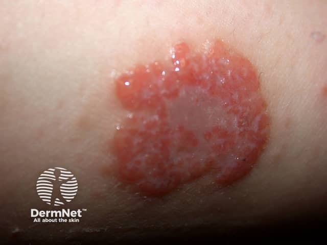
- Subepidermal bullae
- Eczematous or urticarial precursor lesions
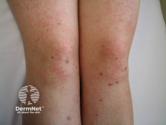
- Associated gluten-sensitive enteropathy
- Intensely itchy; vesicles often removed by scratching leaving erosions
- Symmetrical on scalp, shoulders, elbows, knees, buttocks
Other immunobullous diseases
- Cicatricial pemphigoid
- Pemphigoid gestationis
- Linear IgA dermatosis
- Epidermolysis bullosa acquisita
- Pemphigus vulgaris
- Pemphigus foliaceus
- Paraneoplastic pemphigus
- Drug-induced pemphigus
- IgA pemphigus
- Ocular involvement in autoimmune blistering diseases
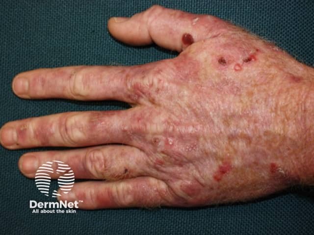
- Metabolic photosensitivity
- Skin fragility, bullae, milia
- Backs of hands, face
- Onset in middle age